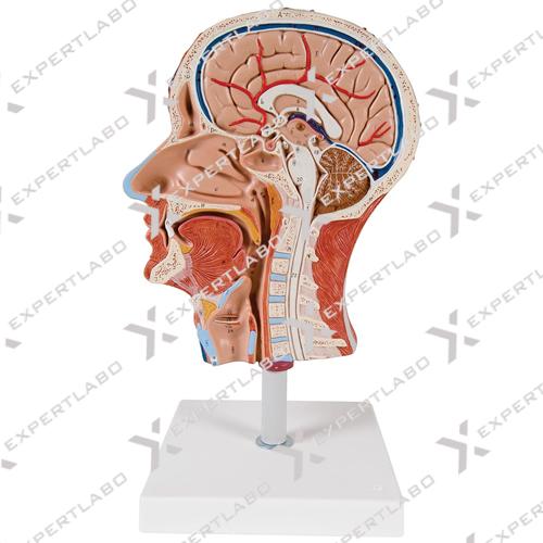
Life size model shows the right half of the human head and neck, sectioned along the sagittal plane. A superficial dissection exposes the facial muscles, the superficial blood vessels and nerve branches of the face ...
View details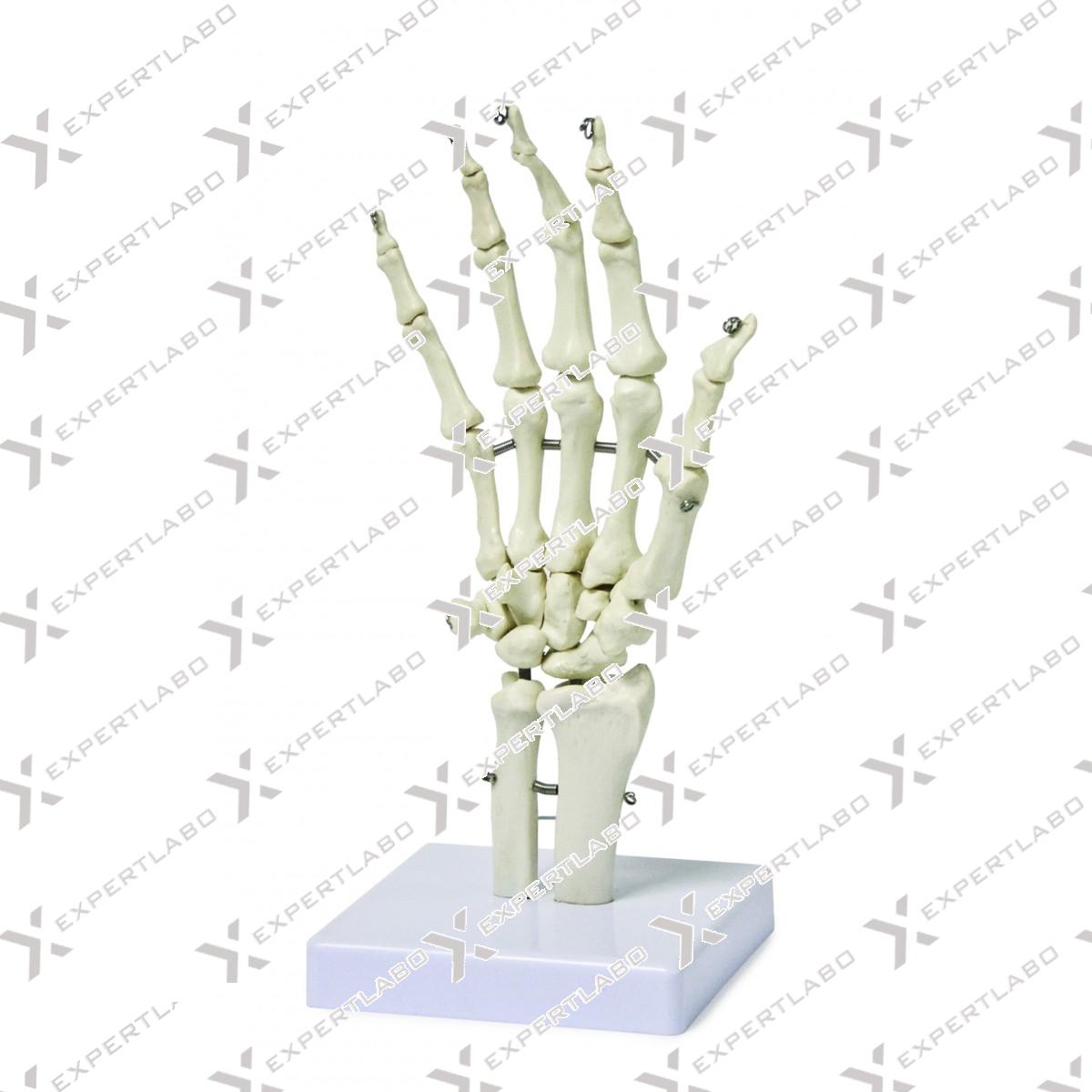
Life-size human hand skeleton model Distal portions of the ulna and radius are present Fully articulated with wire allowing for natural movement Mounted on base Model is made exclusively from high quality medical PVC
View details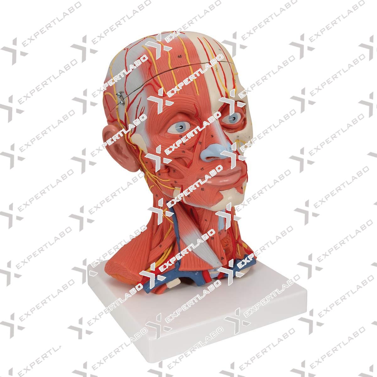
Human head and neck musculature model for anatomical study of superficial and deep muscles, nerves, and vessels Dissembles into five parts, including skull cap and three-part brain, for effective demonstration Mounted on removable base for ...
View details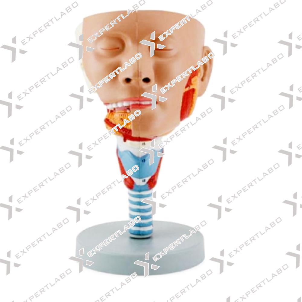
This life size head model is dissected along the sagittal plane into 2 halves. Details of the oronasal cavity and larynx as well as musculature of the pharynx are exceptionally well represented. Mounted on base with ...
View details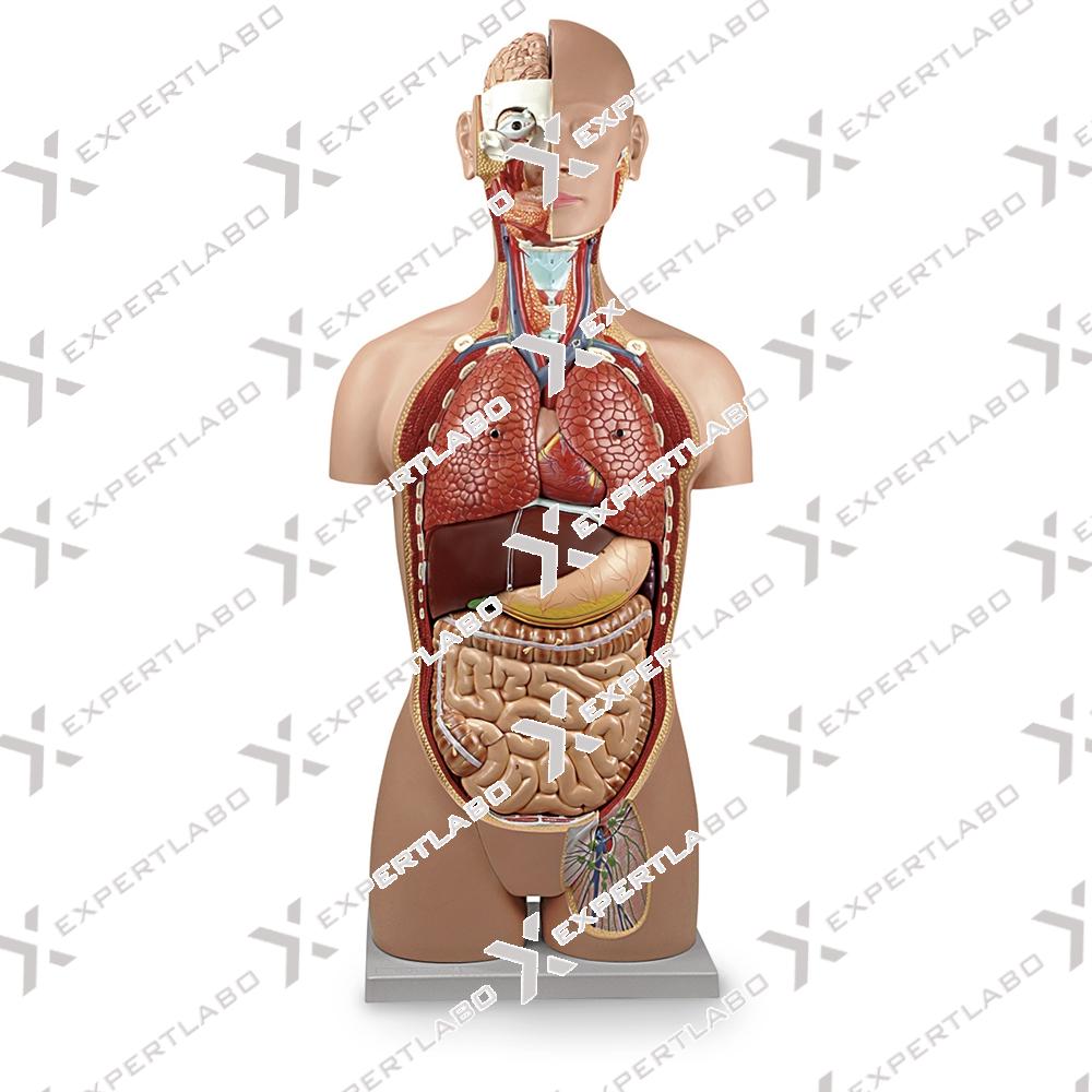
7th thoracic vertebra Removable 6-part head 2 lungs 2-part heart Stomach Liver with gall bladder 2-part intestinal tract Front half of kidney Front half of urinary bladder
View details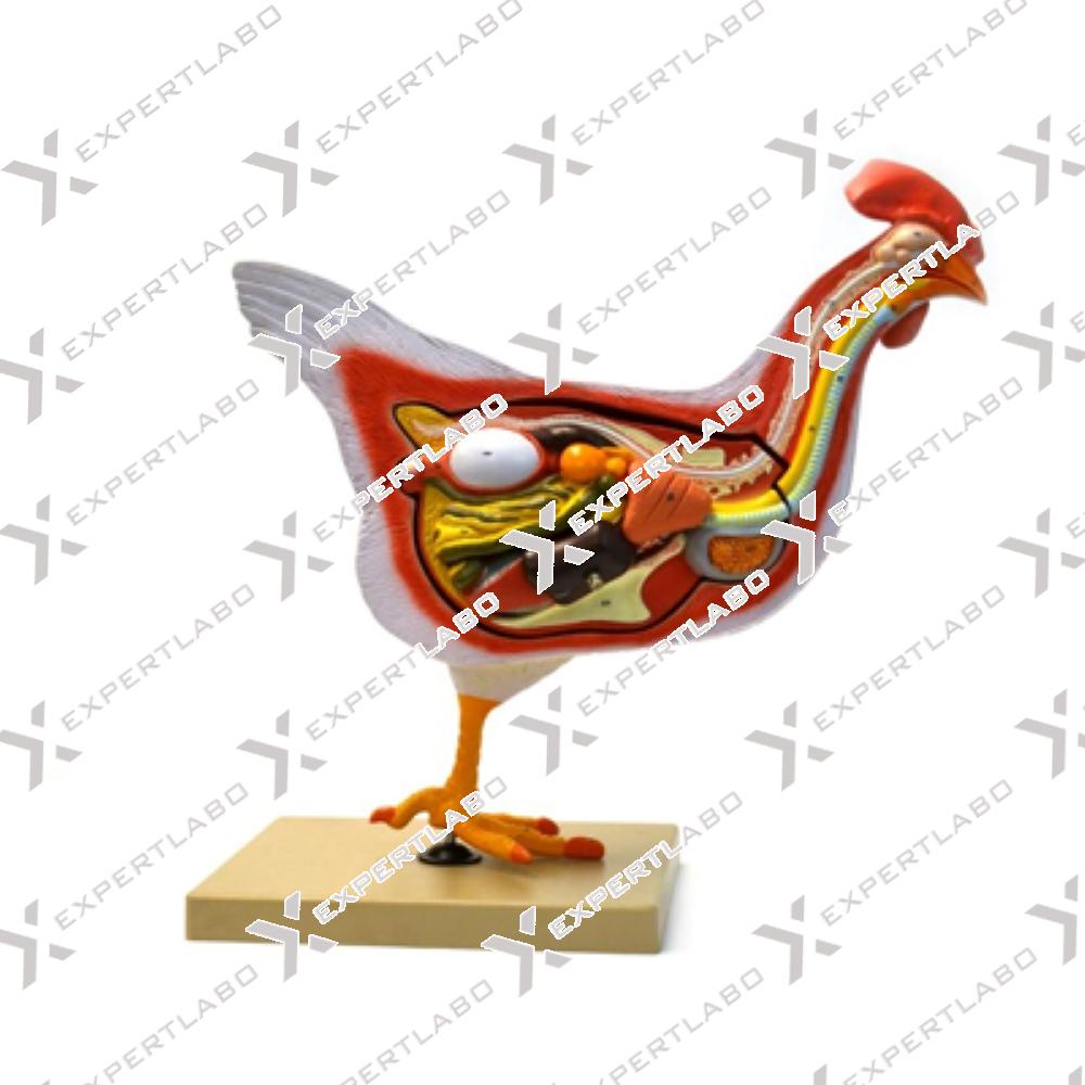
This 6-part life-size model describes in great detail the anatomy of a hen. The right side shows the external morphology while the right part, longitudinally sectioned, reveal the inner structures such as: muscles, heart, liver, ...
View details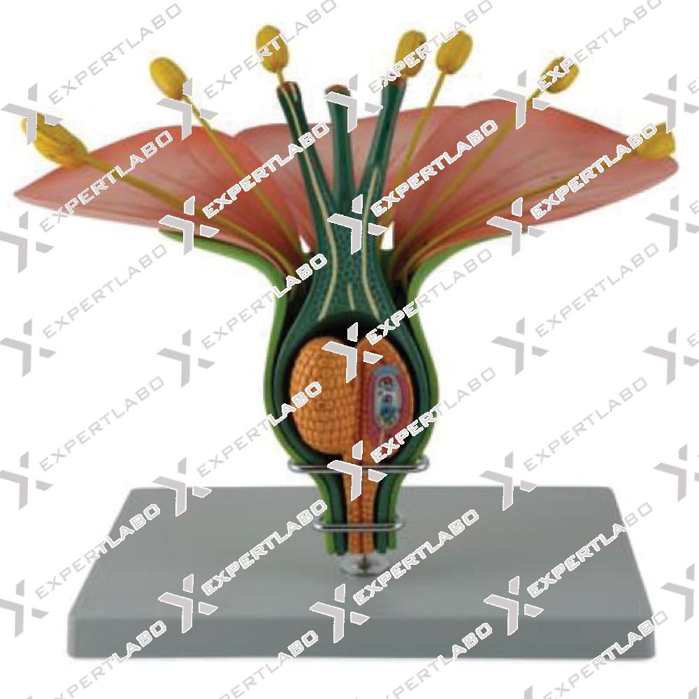
This three-piece model, enlarged to 14X life size, describes with great accuracy and detail the anatomy of a typical hermaphrodite dicotyledonous flower. Thanks to the cross-section, the anatomy of the internal structures, such as stamens, ...
View details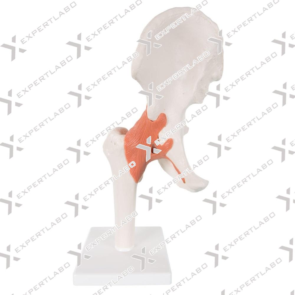
This single-piece life-size model illustrates the right hip joint with ligaments with mounted on board.
View details
This model, enlarged millions of times, shows the structure of the HIV retrovirus, including the outer lipid membrane with protein structures and the internal nucleus containing the viral genome (RNA). This model is supplied with ...
View details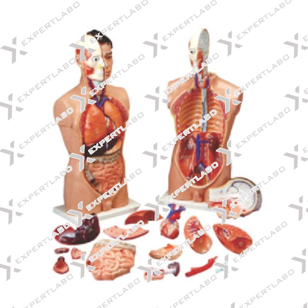
atural size dissectible into 18 parts, mounted on base with key card. Showing ribs, sternum, clavicle with their attachments, thoracic and abdominal organs detachable individually like lungs, heart, stomach, liver, intestine for detailed study. Half of ...
View details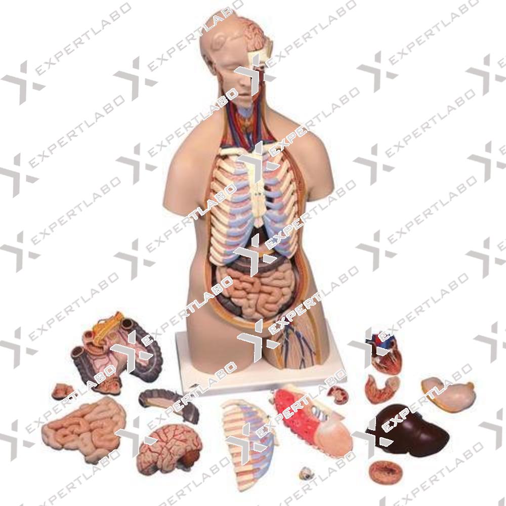
This life size sexless torso mounted on a base, composed of 16 parts, All of the major organ systems are represented with great attention to accuracy and detail. Structures are numbered and identied on the ...
View details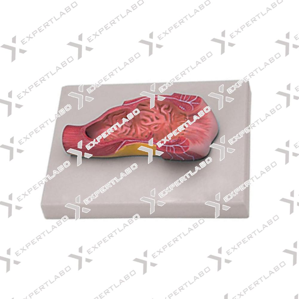
This life size model shows a frontal section of the human anus. Structures of the rectum, including the internal and external sphincter muscles, mucous membrane, ampullae and anal valves are readily visible with mounted on ...
View details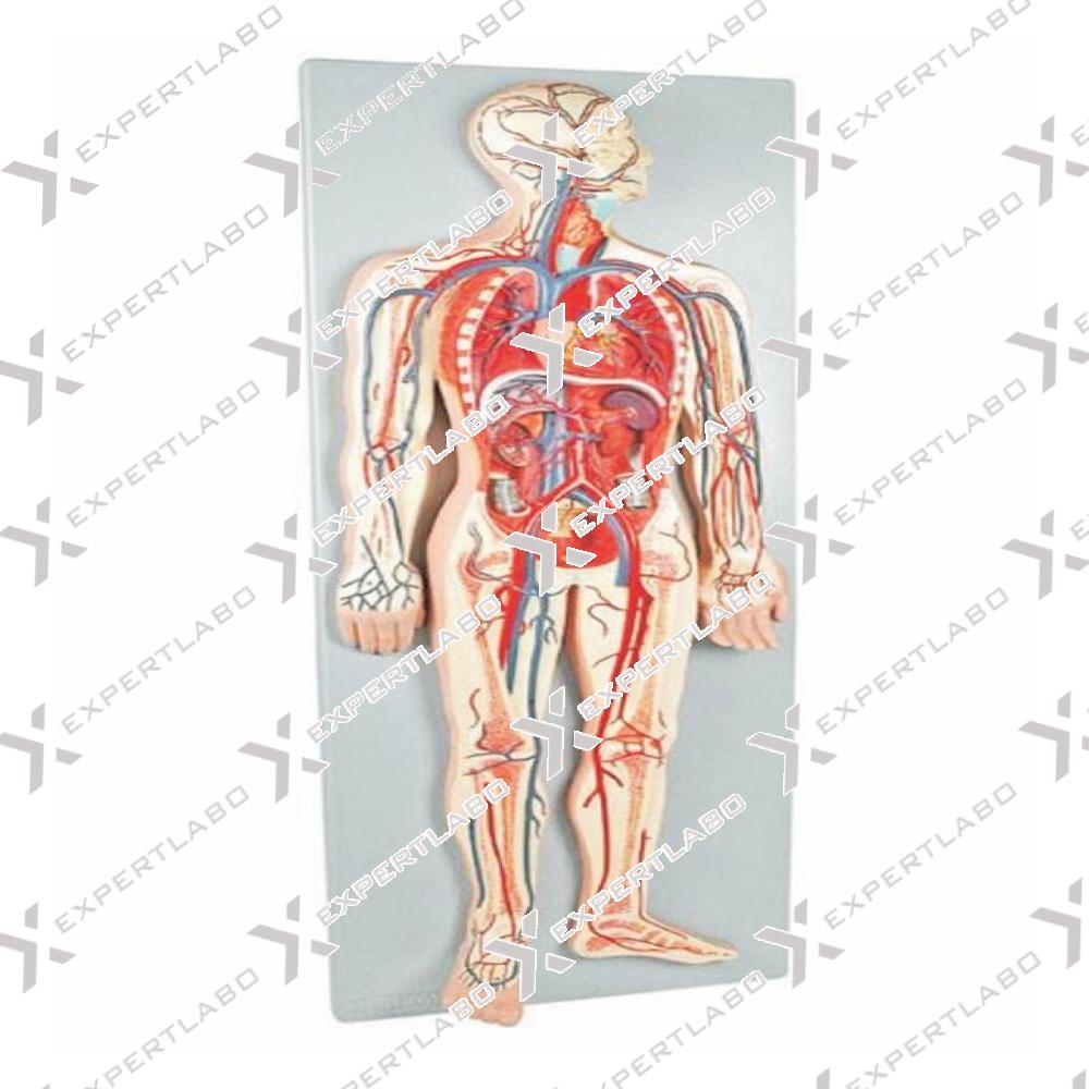
The model represents a general view of the human circulation. It includes the heart (2 parts), lungs, liver, spleen, kidneys and relevant connections with the pulmonary and systemic circulatory pathways with mounted on board.
View details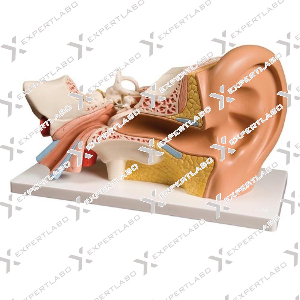
3X life size. The model shows details of the external, middle and inner ear. The eardrum with malleus, incus and stapes are removable. The other removable part is composed of the cochlea and labyrinth with ...
View details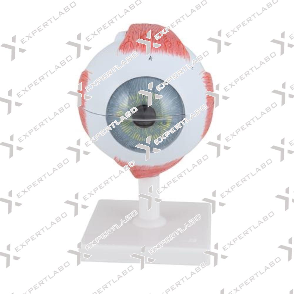
The different parts of the eyeball model are detachable to show the following structures. Tunica External: Showing comea and sclera with attachment of ocular muscles and optic nerve.
View details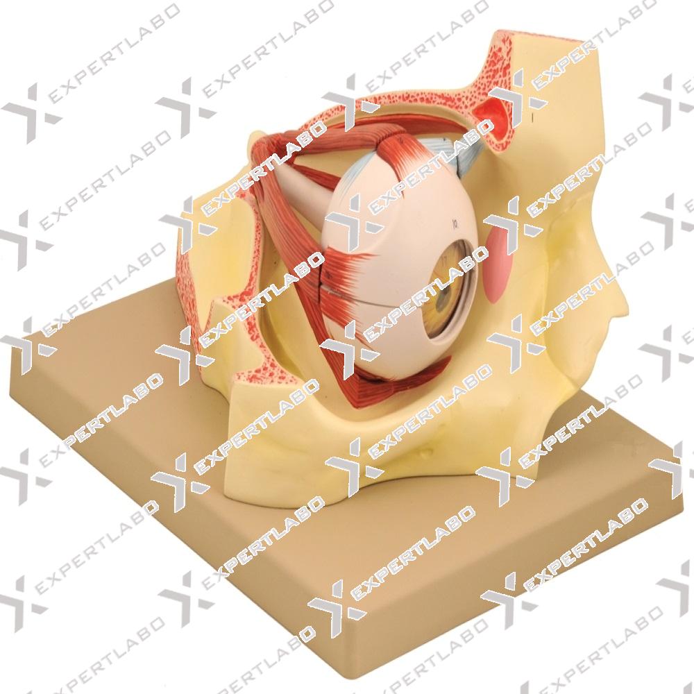
Enlarged 3 times life size, this model includes a dissectible eye and the bony orbit of the skull. The upper half eye ball is removable and dissects into 6 parts showing all anatomical details. Shows the six ...
View details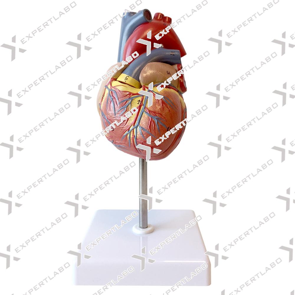
Life size. This 2-piece model shows external and internal anatomy of the heart with origins of pulmonary artery, pulmonary vein, and aorta. Heart valves and chambers are well represented. Includes full-color, illustrated key with mounted ...
View details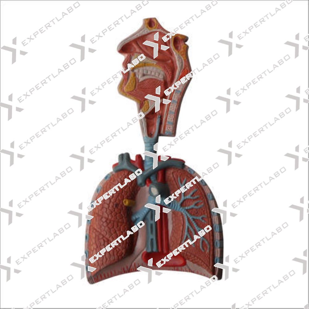
A life size reproduction of the complete human respiratory system. It is composed of 7 parts showing the larynx (dissected along the sagittal plane), the lungs (dissected along the frontal plane) and a 2-part heart with ...
View details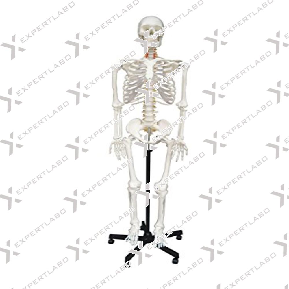
This model is a replica of a life size human skeleton and shows all the skeleton parts in high details. It’s hand assembled to provide intricate details and long-lasting durability. The main joints are ...
View details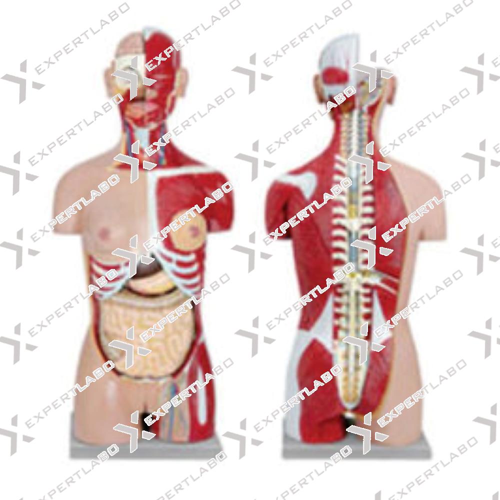
Dissectible in 12 parts, Male/Female reproductive organs can be easily taken out dissectible into 2 parts, without ribs cage. The back depicts the complete vertebral column, spinal cord and nerves. Part of neck and back are ...
View details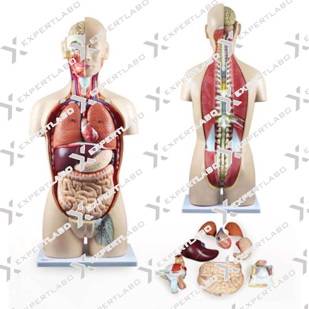
This sexless, life-size torso on base is accurate in all of its detailing. Anatomical structures of the major body systems are numbered an identified on the accompany key. The head is sectioned to expose one ...
View details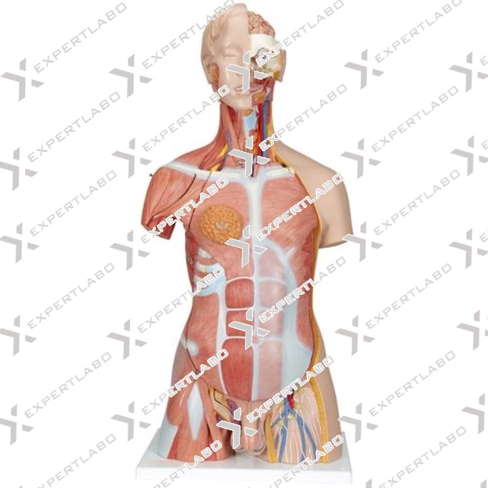
Dissectible in 17 parts, this life size torso with open back is provides an exceptionally realistic reproduction of anatomical structures in their finest detail. All of the body systems are represented with full accessibility. The left ...
View details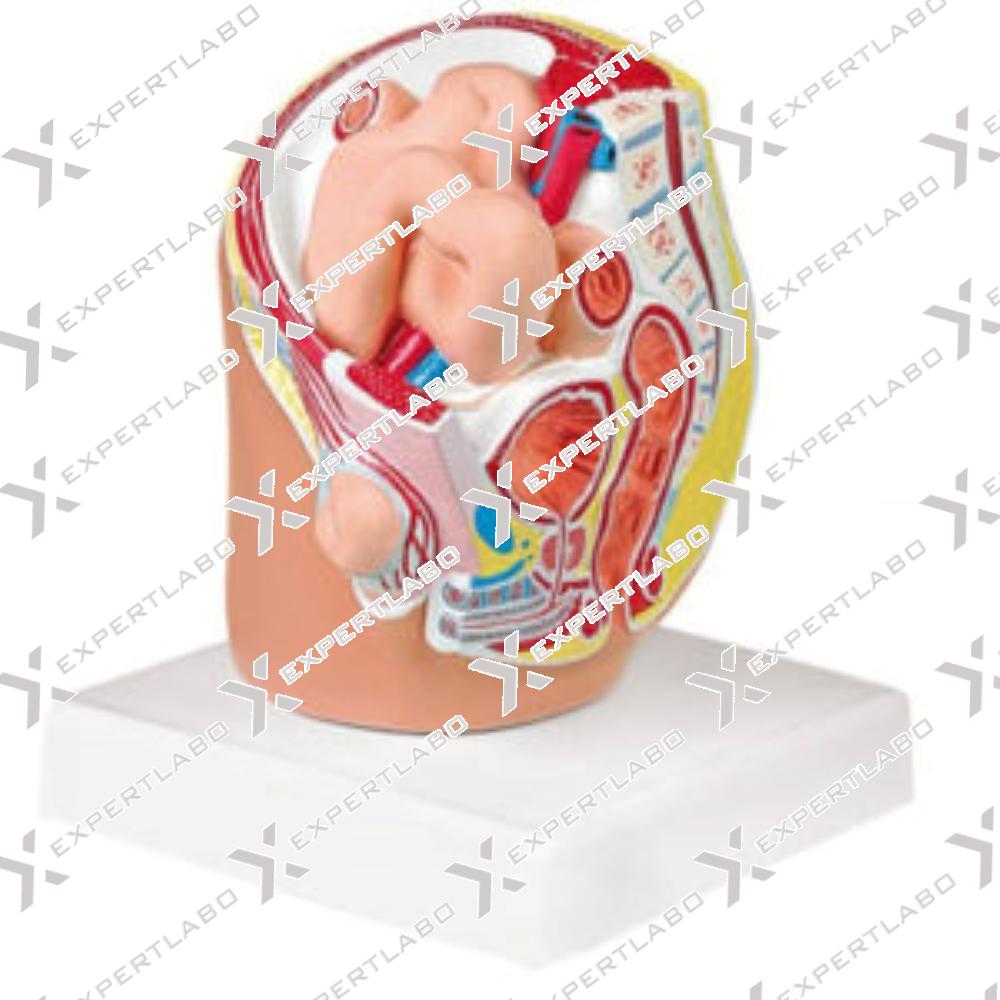
This reduced size pelvis model shows the male internal organs at approximately half life size. The model has been sectioned in the medial and sagittal planes allowing for a clear view of an indirect inguinal ...
View details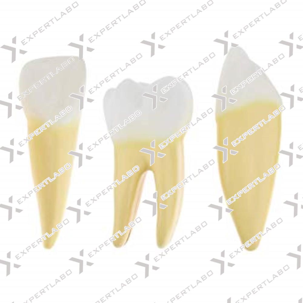
5-part model includes representations (10X life size) of human incisor, canine and molar teeth. The canine and molar can be divided into 2 parts each, showing internal features (dentine, enamel, cement and pulp).
View details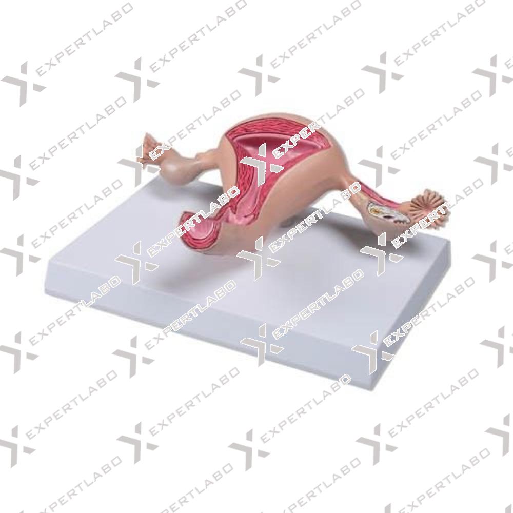
This is a life size model of a healthy uterus. The anatomy is shown in detail, structures are carefully painted by hand. The Cervix, endocervical canal, uterine cavity are sectioned to show endometrium and myomterium. ...
View details
This life model shows the human hyoid bone with mounted on base with stand.
View details/1.jpg)
A life size model of the kidney with the adrenal gland. The model is sectioned along the front plane; it can be divided in 2 parts to show internal structures, including the cortex, medulla, pyramids with ...
View details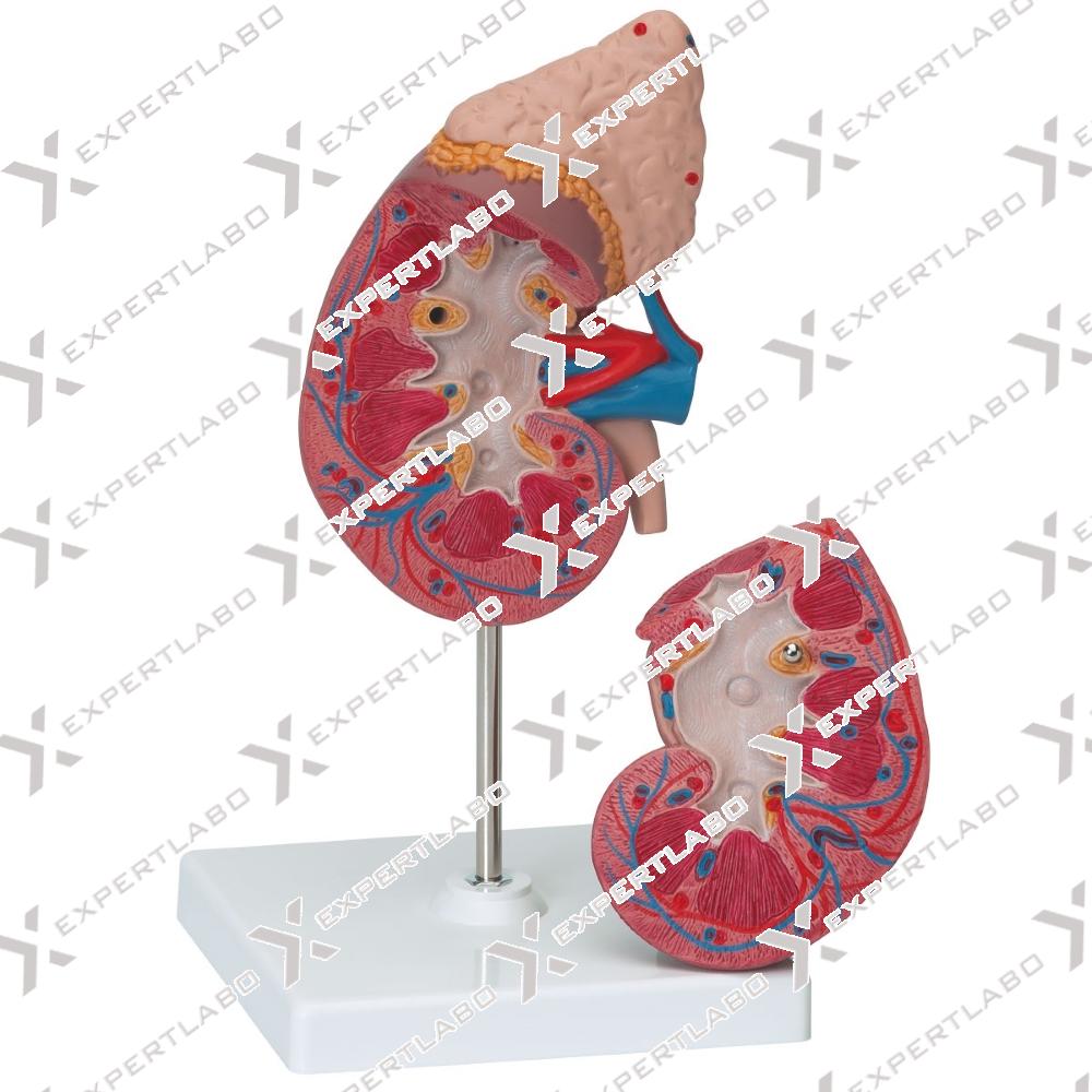
This 2-part model, 1.5X life size, shows the human kidney in frontal region. Internal structures are clearly revealed, including cortex, medulla, pyramids, calyces, renal pelvis (partially opened), ureter and origins of the renal artery and ...
View details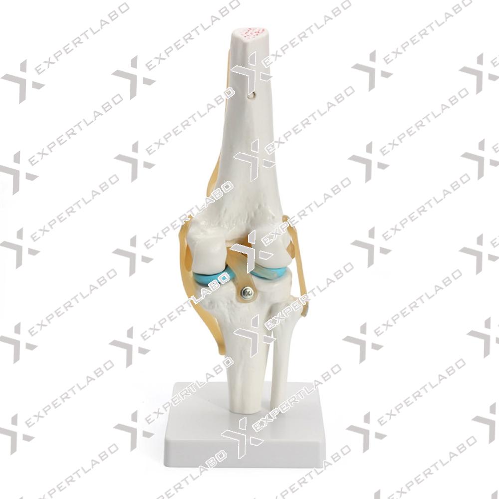
This life-size and fully flexible joint to demonstrate abduction, ante version, retro version, internal/external rotation and much more Consists of portion of femur, tibia and portion of fibula; also includes meniscus, patella with quadriceps ...
View details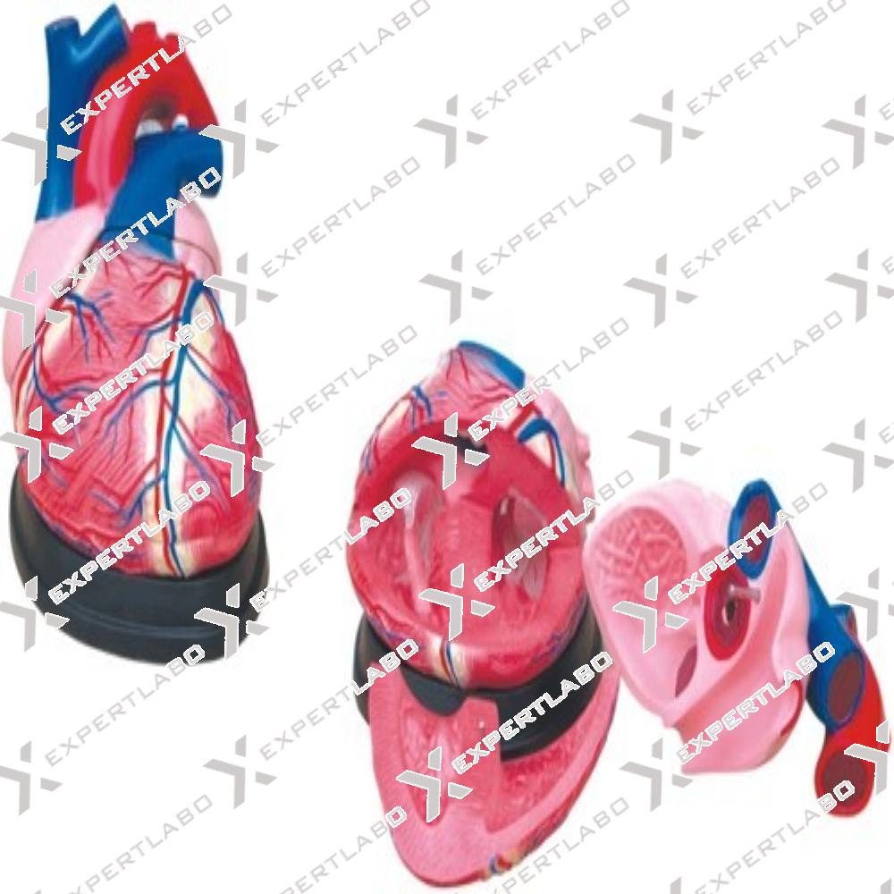
This model shows the external features and internal structures of the heart and its relation with the large blood vessels. A clearer conception of the routes of the systemic and pulmonary circulation can be obtained. ...
View details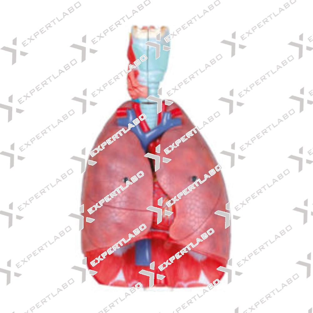
This life-size model separating into 7 parts. The lungs have two removable lobes to show the internal structures. The heart bisects showing atria, ventricles and valves, the larynx bisects and the diaphragm is shown with mounted ...
View details/1.jpg)
This life size model shows a section of the liver with gall bladder, pancreas and duodenum, hepatic and pancreatic ducts with mounted on board.
View details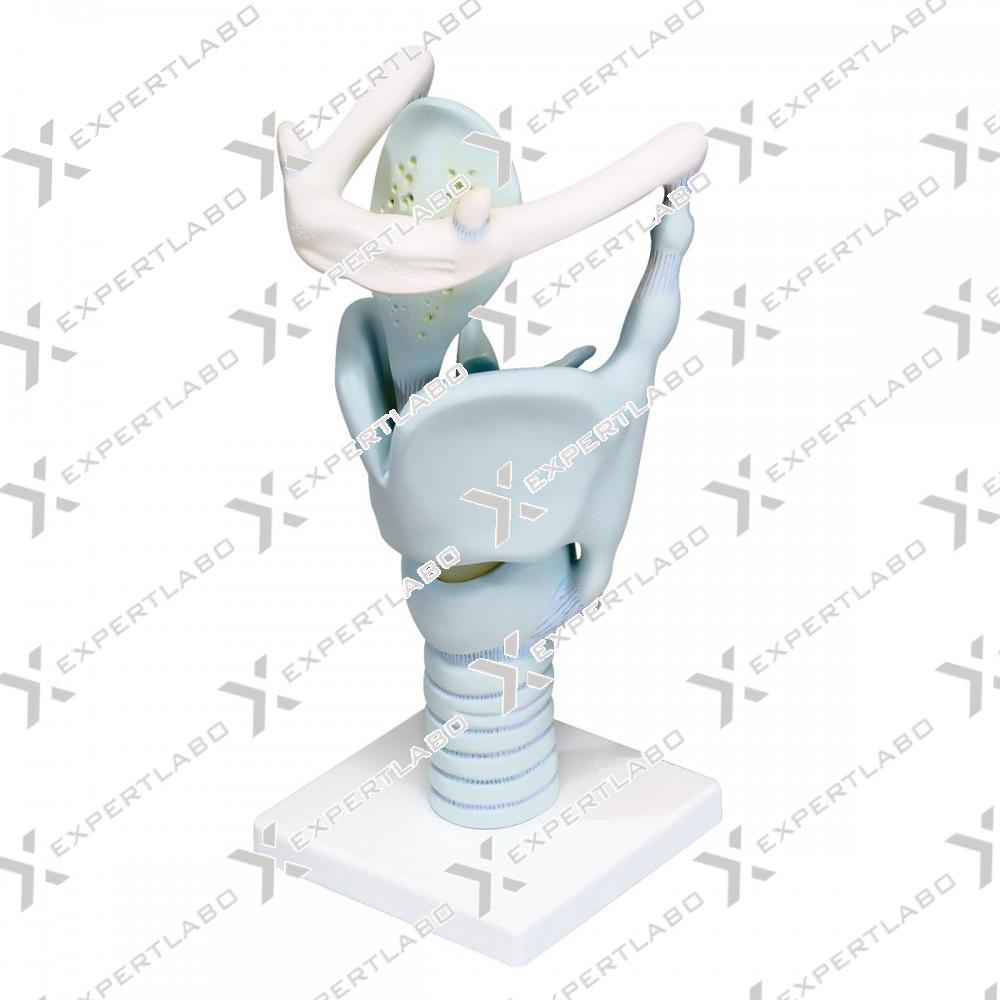
This life size model can be opened into 2 parts to show the anatomy of the human larynx and related structures: thyroid bone, cartilages, ligaments, muscles, vessels, nerves and thyroid gland with mounted on stand.
View details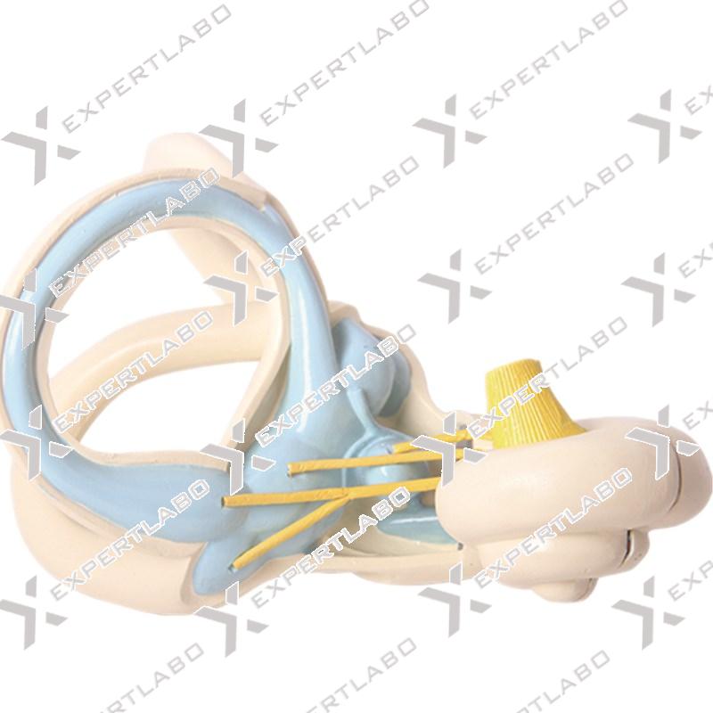
Enlarged about 18 times. Composed of 2 parts. The superior semicircular canal and vestibule are open, Showing the saccule and utricle.
View details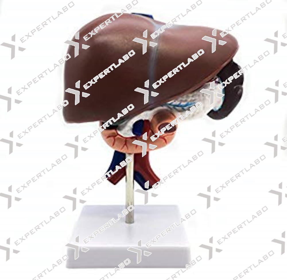
This realistic model reproduces a life size liver with the gall bladder. The hilus vessels are shown as well as the extra hepatic ducts and the main ligaments. Mounted on base with stand.
View details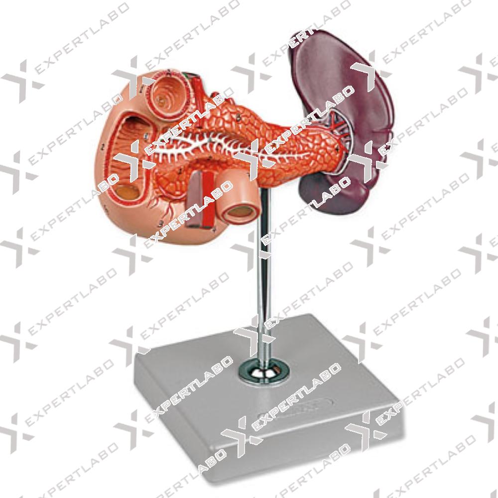
This life size model (2-part model) shows the liver with gallbladder, pancreas and partly dissected duodenum. It includes inferior vena cava, abdominal aorta and pancreatic ducts. Mounted on base with stand.
View details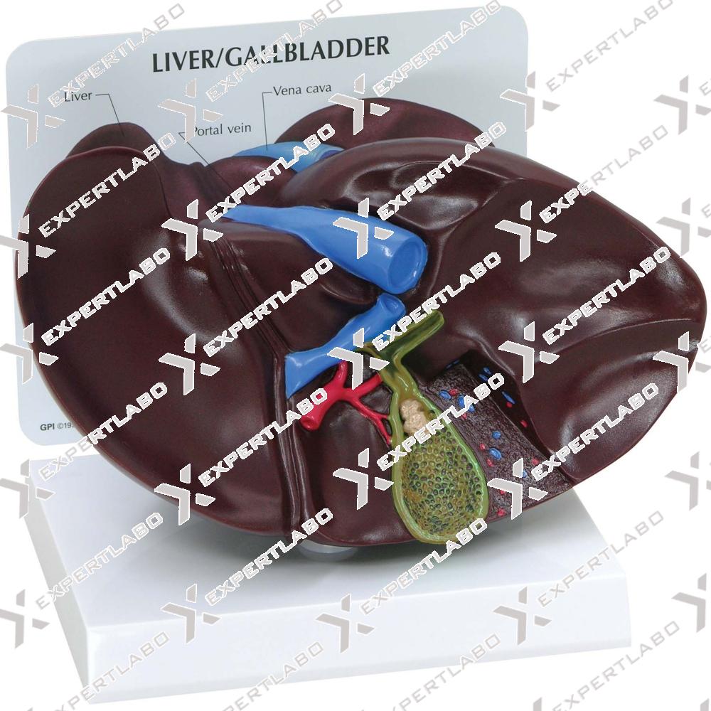
The model, 1.5X life size, shows the liver and gallbladder in open section with the complex network of vessels and bile ducts, displayed in different colors for clarity and ease of examination. Mounted on base ...
View details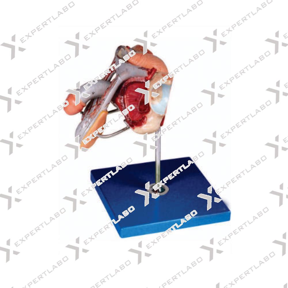
This life size model, composed of 4 parts, is dissected through the median sagittal plane to provide excellent views of external and internal structures. The removable parts include two halves of the penis, showing medial and ...
View details/1.jpg)
This life size model, composed of 4 parts, presents an open view through a median sagittal section of the pelvis. Internal structures of the male urogenital system are represented in detail. The removable parts include a ...
View details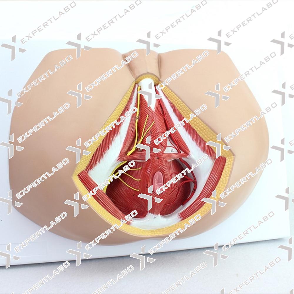
This life size model shows the male perineal area, including the anus and external genitalia. The pelvic diaphragm, urogenital perineum and anal perineum are well represented, including distribution of blood vessels and nerve endings.
View details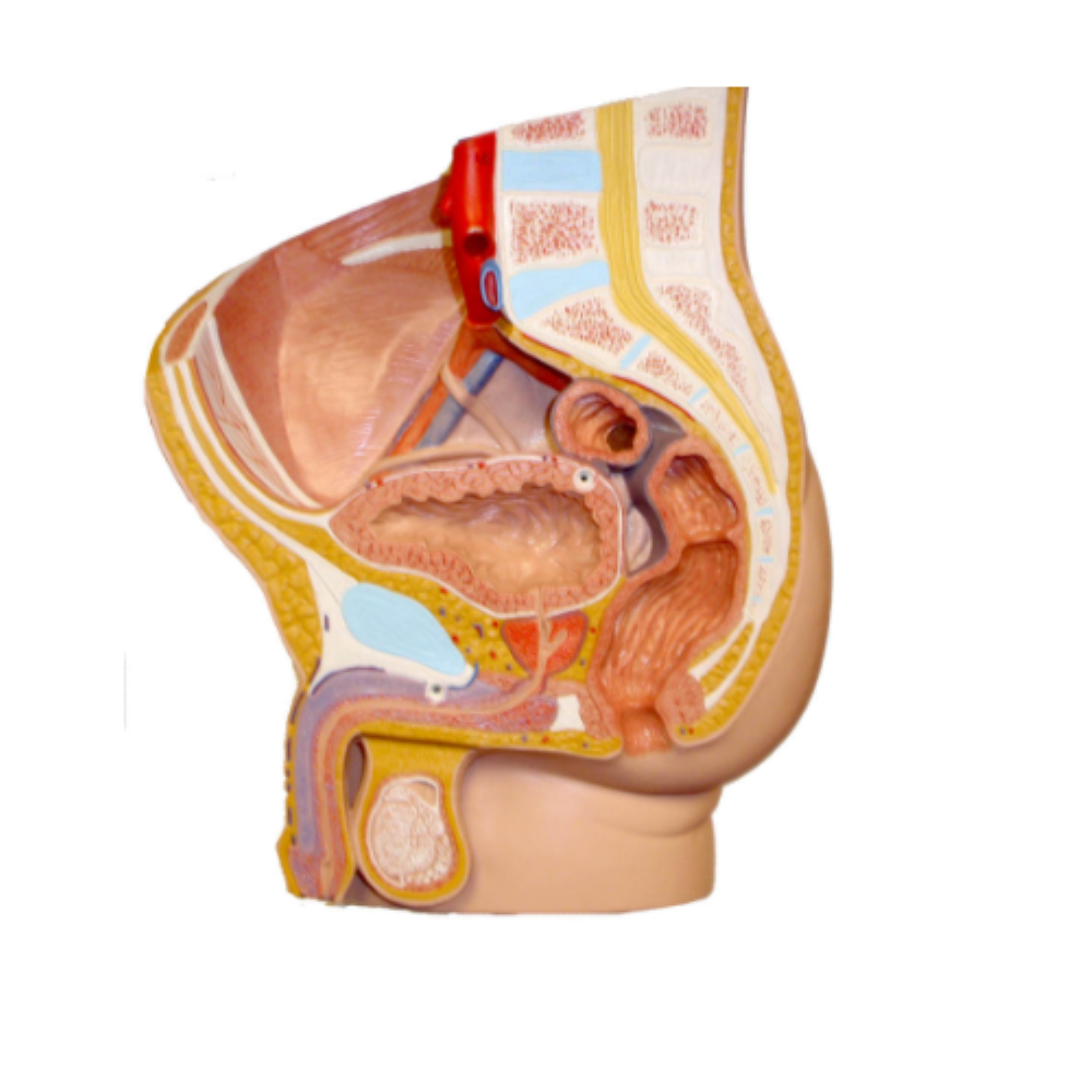
This model shows in natural size, all the organs of the male reproductive system. Penis, prostate and urinary bladder are longitudinally sectioned to reveal their inner anatomy; the testicle is partially cut away to expose ...
View details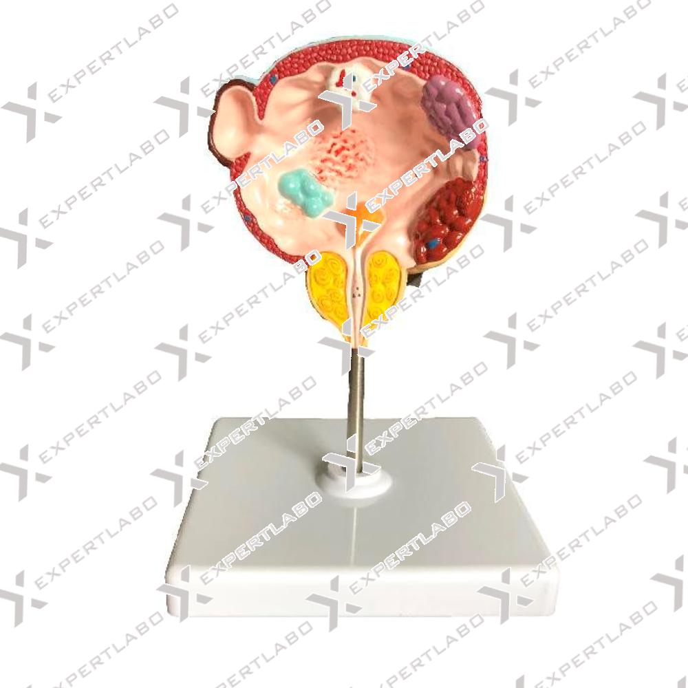
This model, enlarged 3 times, shows the male urinary bladder with the prostate gland surrounding the urethra. The model is dissected medially to expose both internal and external structures of the bladder and prostate, including the ...
View details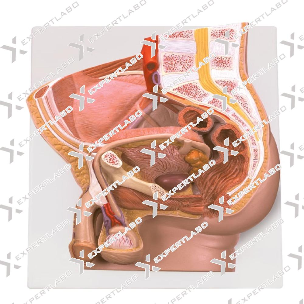
This life size 2-part model shows the internal structure of the urogenital male system with specific pathologies, including gallstones, diverticulum, cancer of the urinary bladder and sexually transmitted diseases such as condyloma acuminatum and gonorrhoea. ...
View details/1.jpg)
Life size representation of the superficial and the internal structures of the head and neck. This relief model shows all relevant structures of the human head in great detail. It’s delivered on a baseboard ...
View details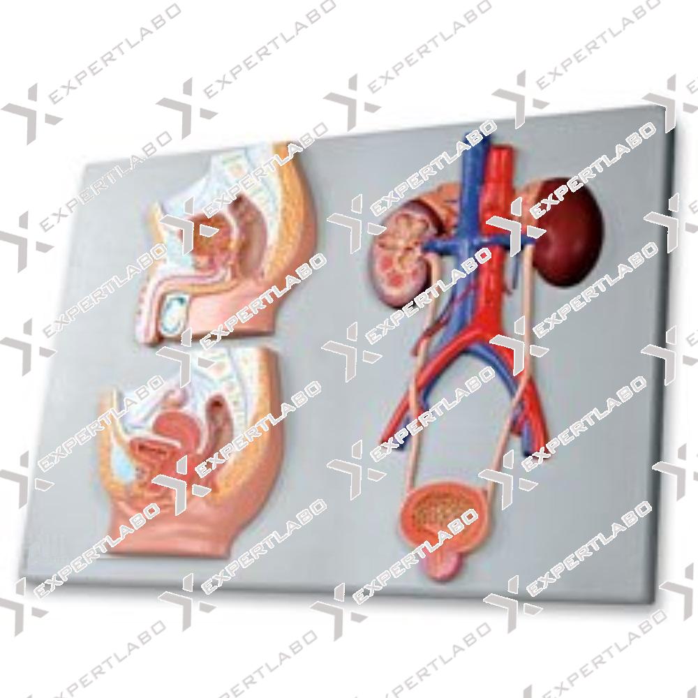
This model features sections of both male and female pelvises, plus the urinary bladder, urethra, the inferior vena cava with tributaries, the abdominal aorta with branches, ureters, the kidneys with adrenal glands, a section showing ...
View details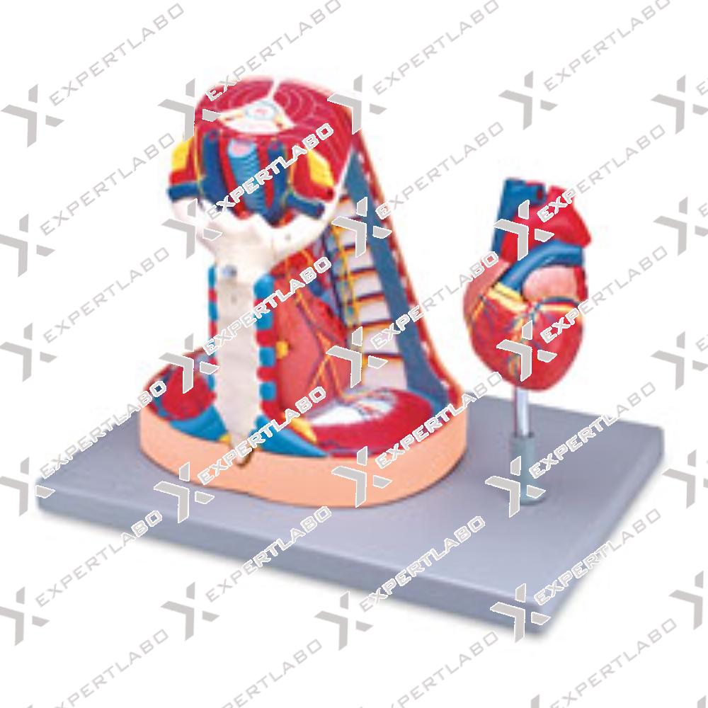
This life size model is composed of 6 parts, including a 2-part heart that provides an interior view of the chambers and valves. The sternum and thymus are removable to reveal the pericardial sac and the ...
View details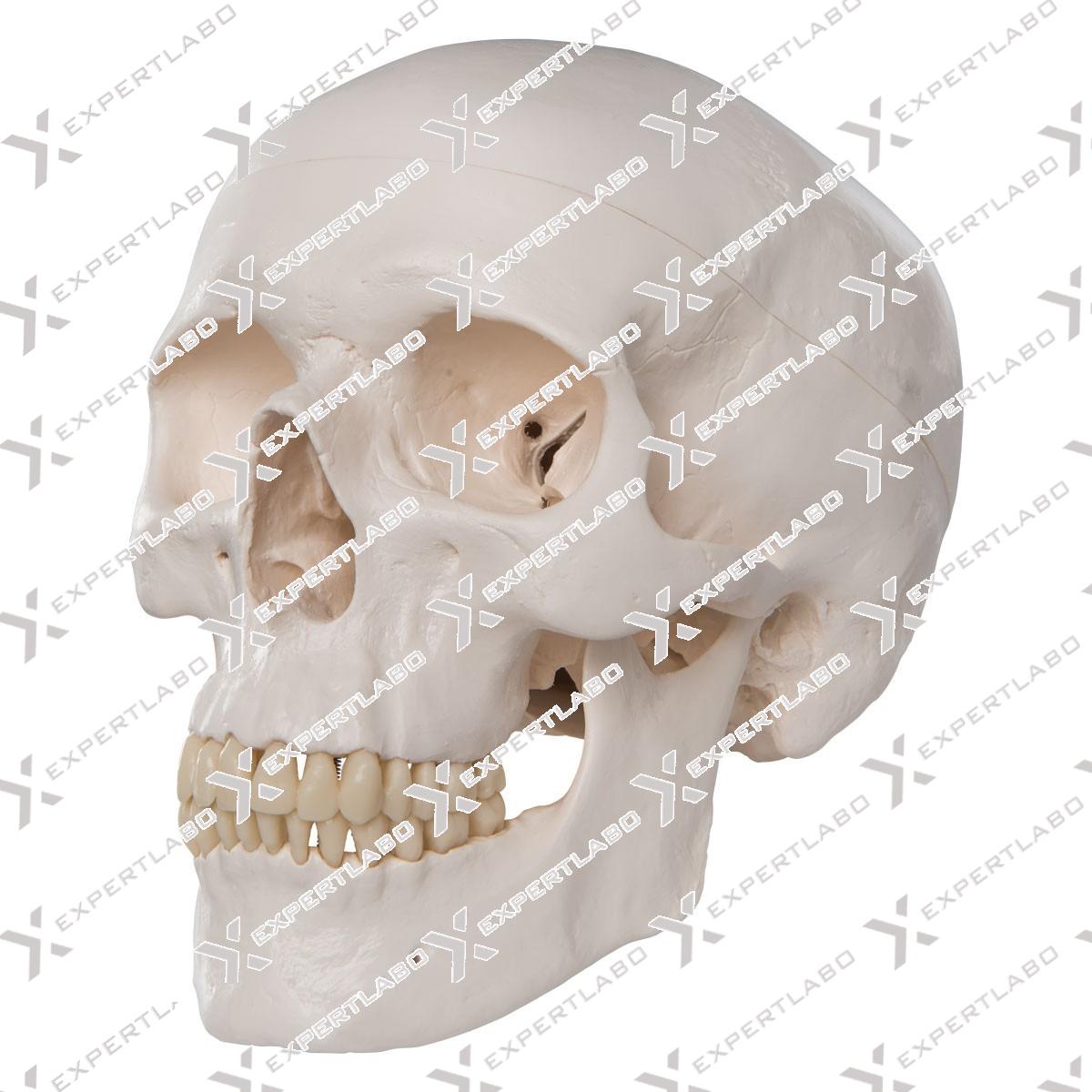
This model, ½ adult natural size, depicts all structural details of the human skull: it can be disassembled into 3 parts: skullcap, base of skull and mandible.
View details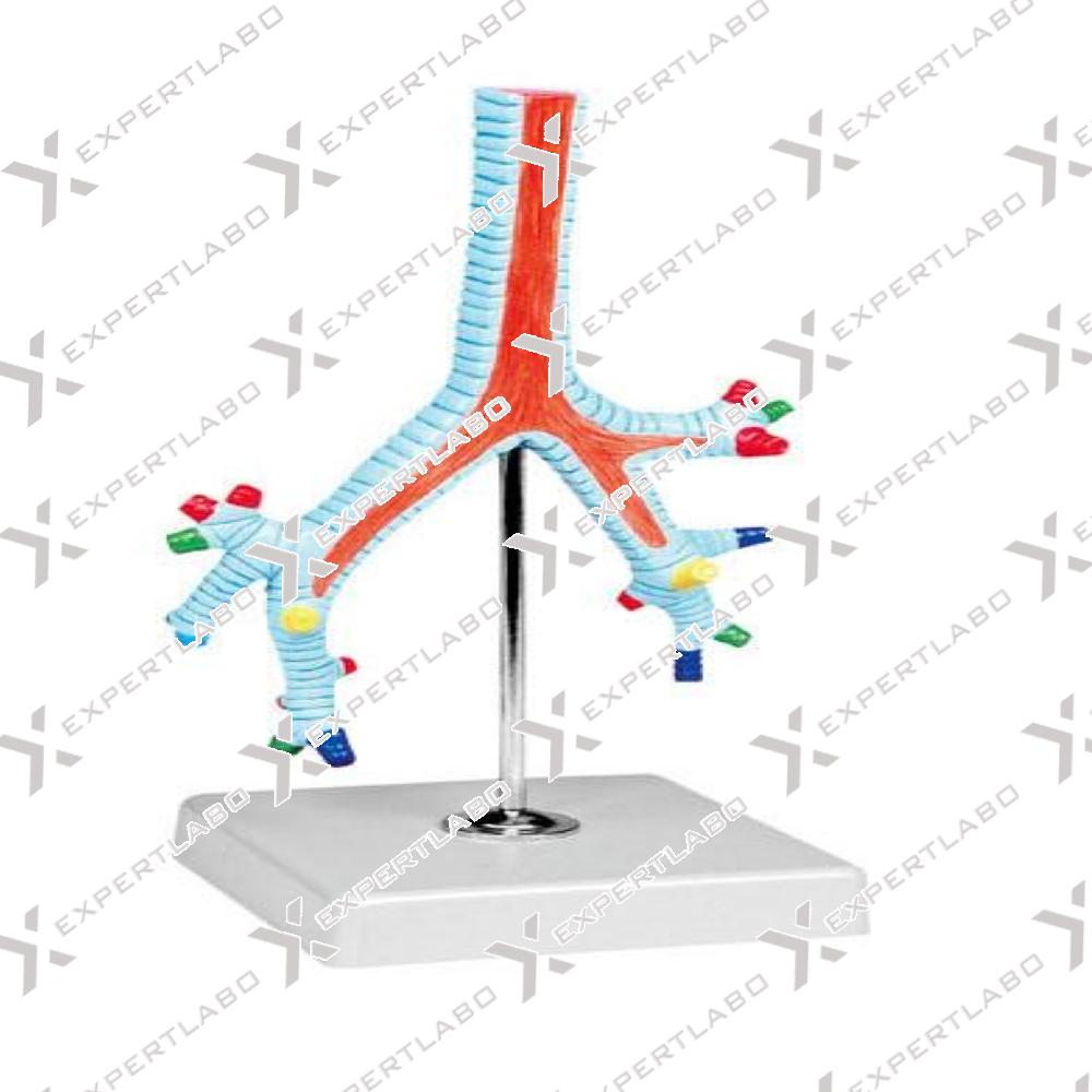
This life size model shows the bronchial tree from the bifurcation of the trachea through its subsequent subdivisions to the bronchopulmonary segments, displayed in different colors with mounted on stand.
View details/1.jpg)
This model illustrates in great details the muscles, ligament and bones of the shoulder. Through different muscles section it is possible to observe the profound musculature as far as to get bone. Life size model ...
View details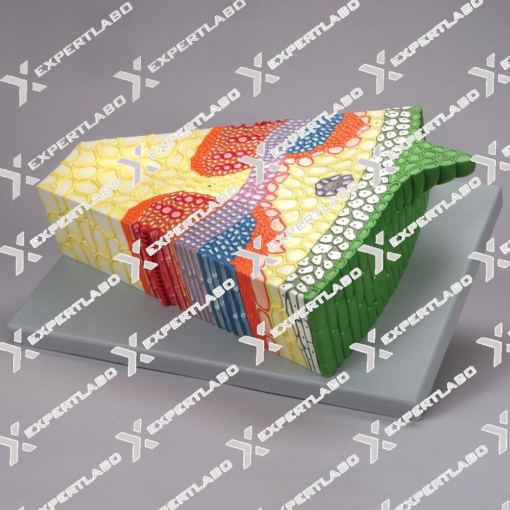
This model, enlarged 500X life size, shows the cross and longitudinal section of a monocot leaf. Significant structures include dorsal and ventral page, xylem, phloem and mesophyll with mounted on board.
View details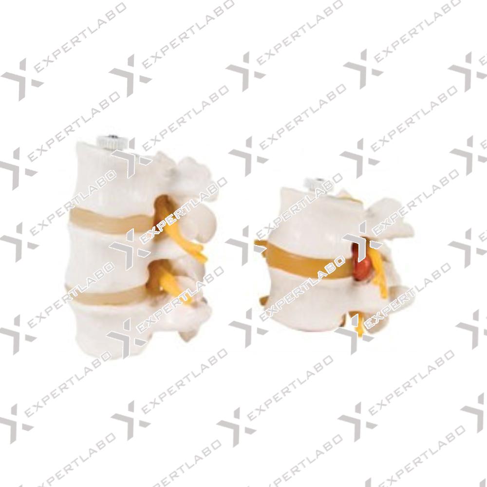
This life size model shows 2 lumbar vertebrae with spinal nerves, dura mater and an inter vertebral disc affected by medial hernia. On the base it shows 2 pathological inter vertebral discs with the correct position of ...
View details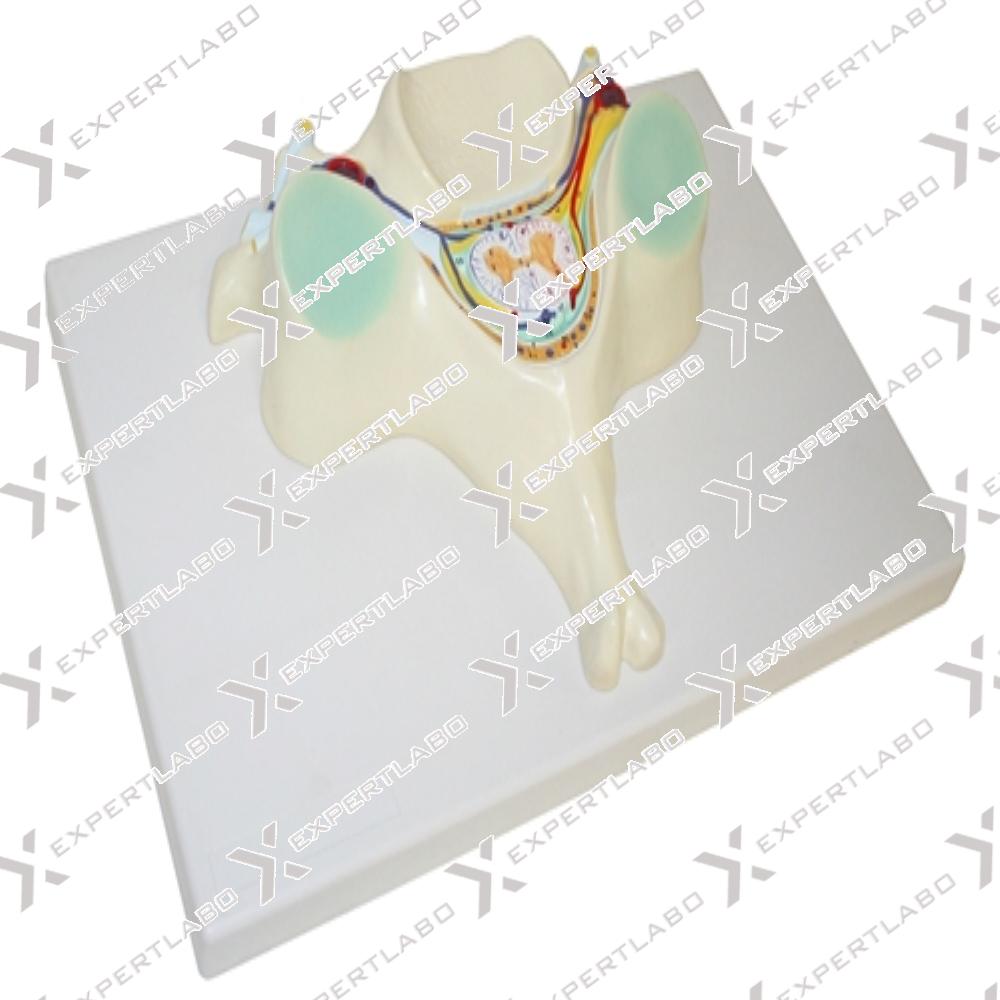
7X life size. This model shows in great detail the structure of the 5th cervical vertebra. a transverse section of the spinal cord with grey and white matter, the nerve branches, the spinal ganglion and ...
View details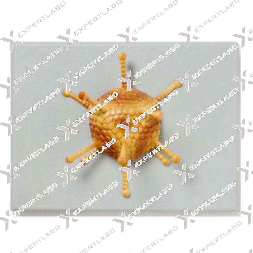
This model shows the caps-id structure of the adenovirus, enlarged millions of times with mounted on board.
View details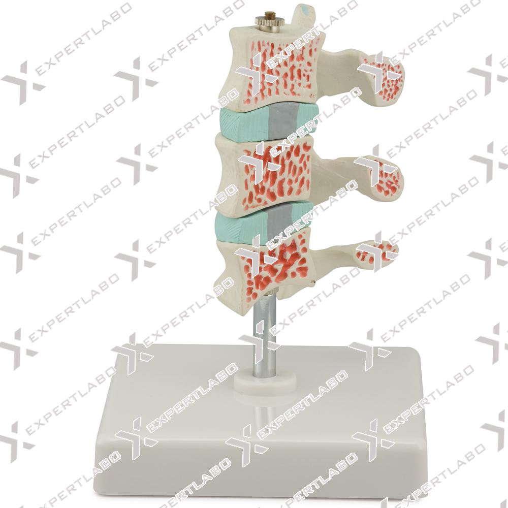
This life size model shows three vertebrae: one healthy vertebra, one vertebra with pathological changes at first stage, one vertebra with pathological changes at advance stage. Mounted on base with stand.
View details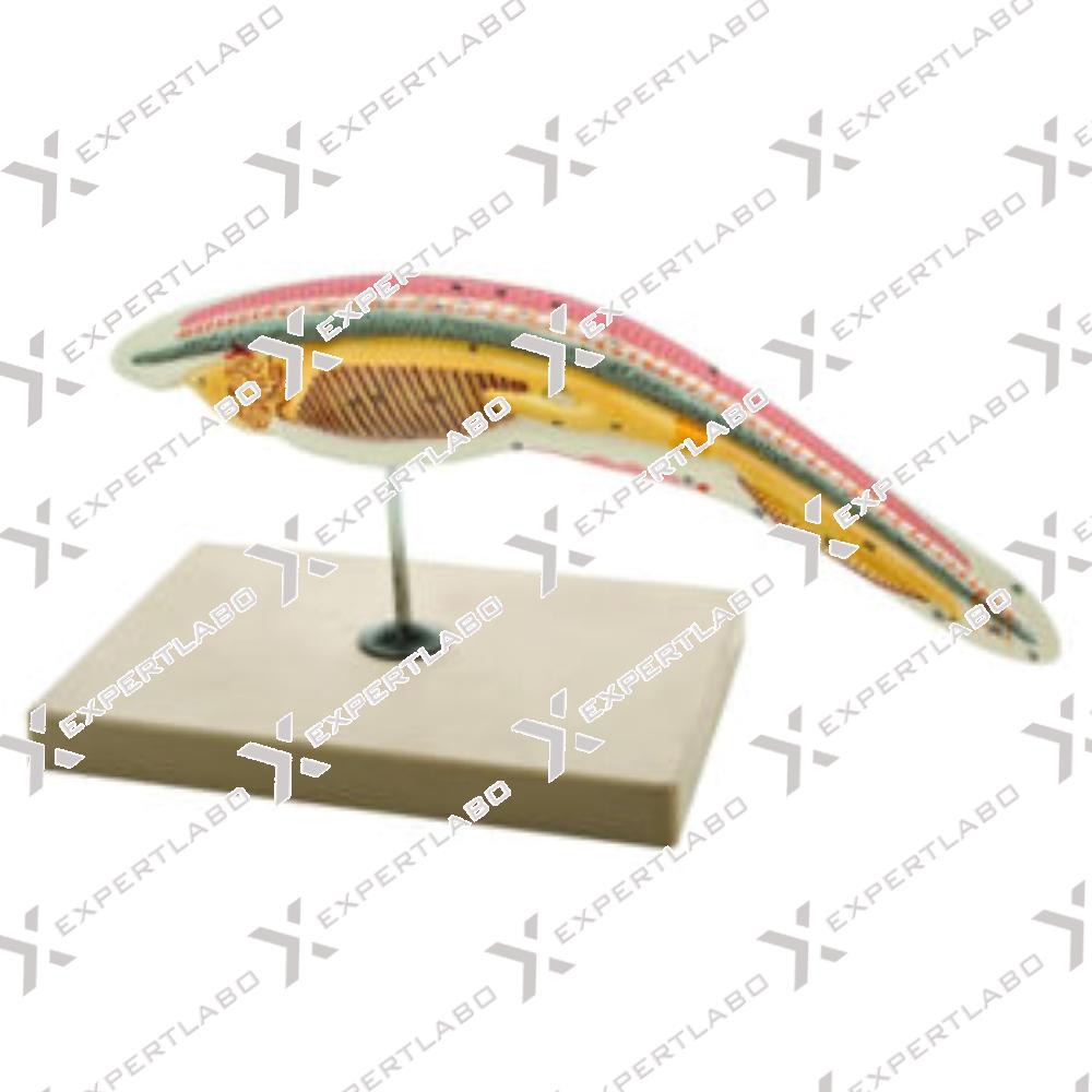
This three-part model, enlarged to 10X life-size, shows the morphology of an Ampioxus. The inner anatomy can be easily studied thanks to the longitudinal section; all the main structures, such as cerebral vesicle, neural tube, ...
View details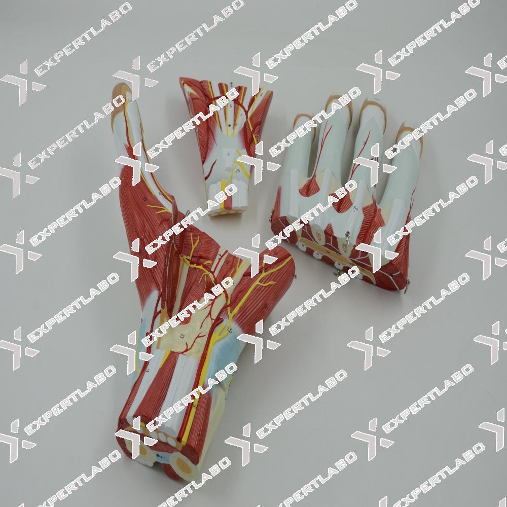
This 3-part model, enlarged 2X life size, shows the anatomy of the hand, including muscles, nerves, ligaments, vessels and bone structures. Aponeurosis is removable for closer examination
View details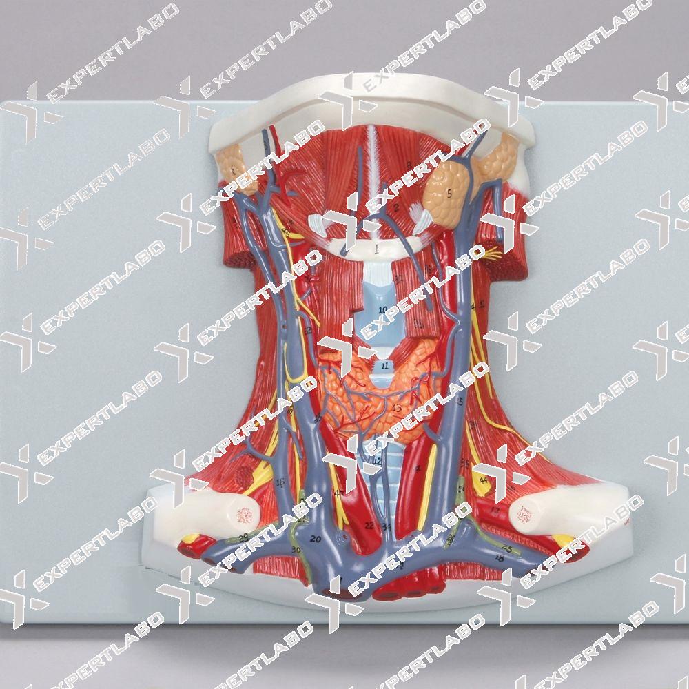
Skin Cancer Model, it’s easy to show three cancerous conditions: NBC(Nodular Basal Cell), MM (Malignant Melanoma), MBC (Morphemic Basal Cell) and three non-cancerous conditions: DN (Dysplastic Nevi), KA(Keratoaconthoma),AK (Actinic Keratosis). The ...
View details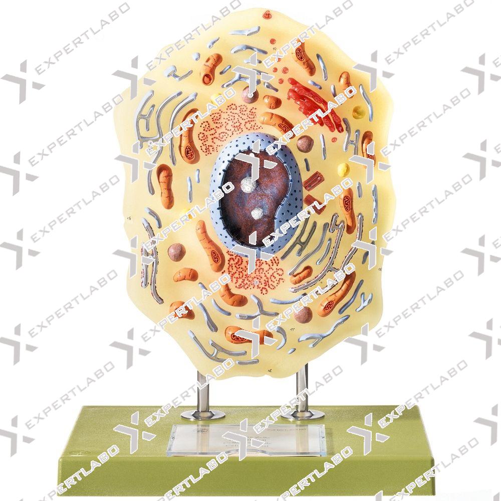
This single-piece model, magnified approximately 13,000 time is a very useful tool to study the cytology of an eukaryotic cell. The typical structures, such as micro villi, flagellum, mitochondria, nucleus, rough and smooth ER are reproduced ...
View details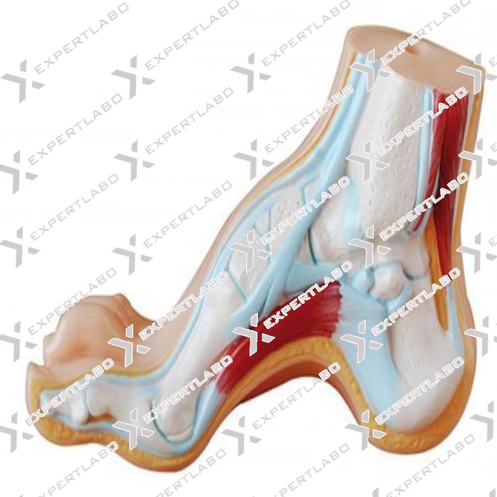
This model shows the anatomical structure and the distal end of tibia of arched foot or flat foot.
View details
Adetailed, life size reproduction of the atlas and axis vertebrae, including the occipital bone. The model shows the pivoting action of the head and the articulation with the skull. Mounted on base with stand.
View details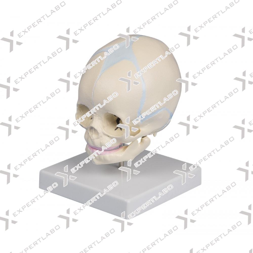
Baby skull is a natural cast of a fetal head in the 30th week of pregnancy showing the characteristics of prenatal development. The fontanel, which becomes bone over time, are clearly visible on the skull. ...
View details
The structure of bacteriophage T4, a virus that attaches to the surface of the much larger bacterium Escherichia coli, is shown millions of times enlarged life size with mounted on board.
View details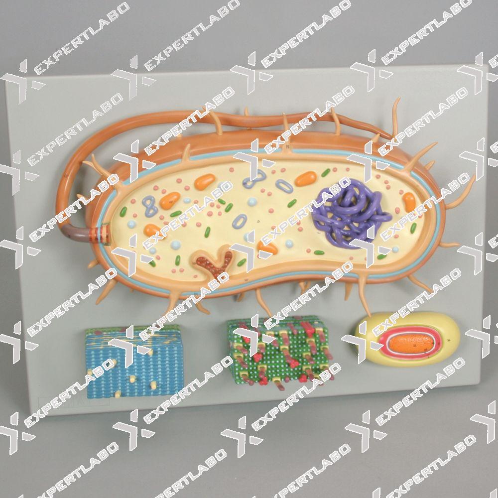
A complete tool for understanding the structure of the prokaryotic cell. This item, greatly enlarged, is composed of four models on the same base: A cross-section of a general prokaryotic cell, illustrating typical structures as ...
View details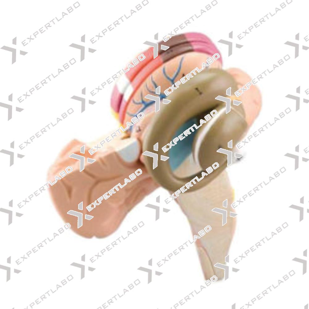
A fundamental support to study the human deep neuroanatomy. Greatly enlarged, composed of 2 parts, it clearly shows – with the aid of false colours – the principal anatomical structures such as the caudate nucleus, the lentiform nucles ...
View details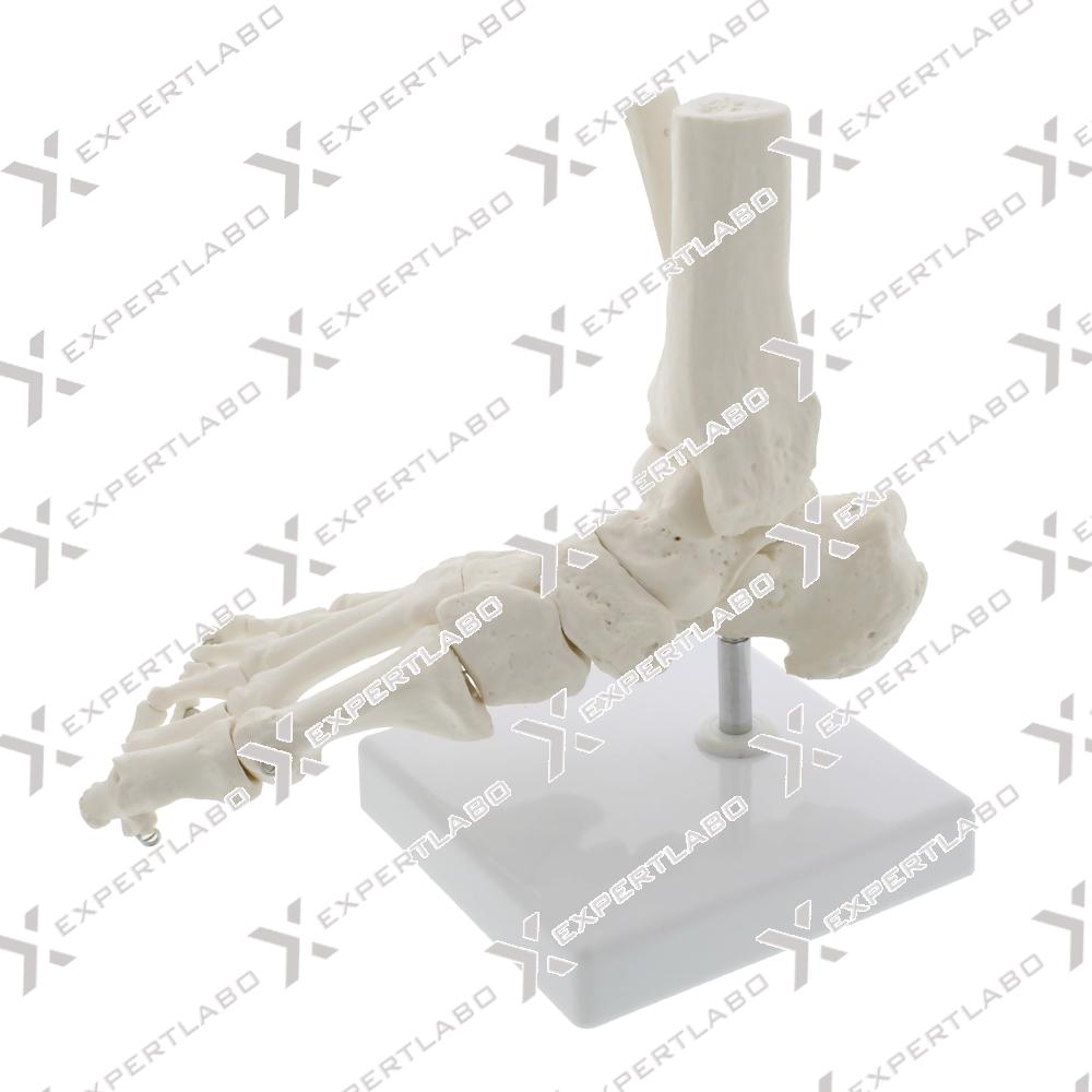
A life size skeleton of the human foot with articulated joints. Natural movements of the foot can be demonstrated easily and effectively.
View details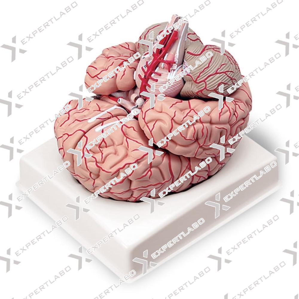
A life size brain model divided medially in 2 parts along the sagittal plane. It shows the left and right cerebrum, cerebellum, brain stem and blood vessels. Includes cradle base.
View details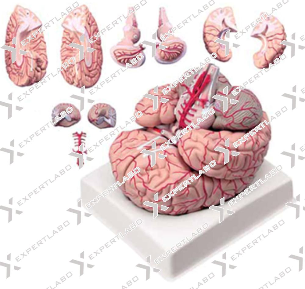
Life size brain with distinct color coding for the following regions: frontal lobe, parietal lobe, occipital lobe, temporal lobe, motor cortex, somatosensory cortex, limbic cortex, cerebellum, brain stem. Mounted on base
View details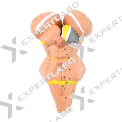
This 4-part model, 2X life-size, is a detailed representation of the human brain stem and hypothalamus. The brain stem is sectioned to show the hypothalami c nuclei; the thalamus is removable and divisible into two ...
View details
4X life size. Human cerebellum is dissected to show details of internal organization.
View details/1.jpg)
This life-size model can be divided into three parts: Cerebellum and Brainstem, longitudinally sectioned, showing the insertions of the cranial nerves and the inner anatomy. All the different structures are numbered and identified on the ...
View details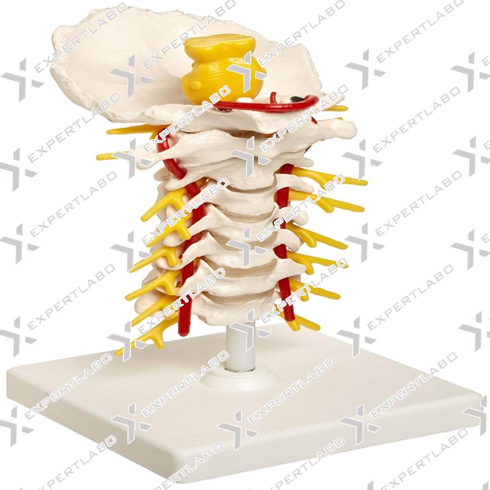
This life size model of the cervical spinal column consists of 7 cervical vertebrae with inter vertebral discs, occipital plate, cervical nerves, vertebral arteries, spinal cord and brain stem. Mounted on base with stand.
View details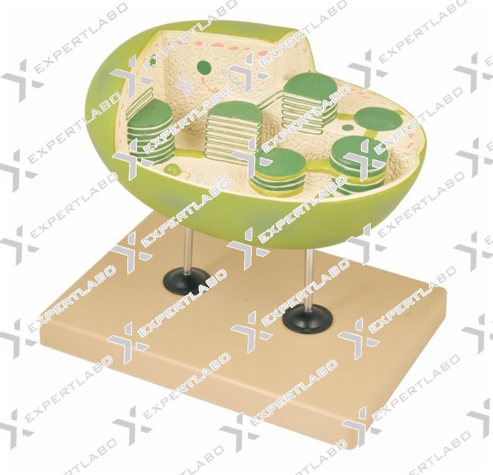
This model represents an important tool to study one of the most important organules of the plant cell. The chloroplast, greatly enlarged, is cross and longitudinally sectioned to show in great detail the internal microscopical ...
View details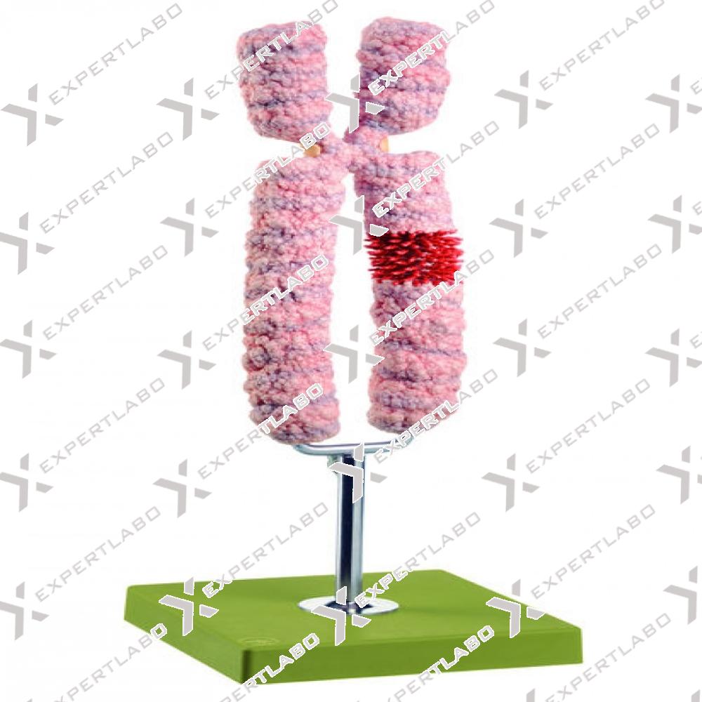
This single-piece model, 10000X enlarged, shows the structure of a human chromosome. All the main parts – centromere, telomere and loops – are extremely accurately reproduced. Mounted on base with stand.
View details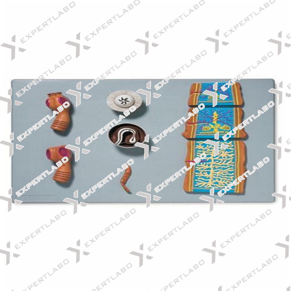
This model compares the structures of Taenia saginata and Taenia solium scolex; the anatomy of the proglottid is shown through the longitudinal section at three different stages of maturation: immature, mature and gravid proglottid. Significant ...
View details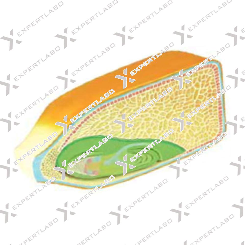
This model is 40X life size; it shows the corn seed in longitudinal section with excellent details of the seed coat and endosperm. The seed embryo can be removed for closer examination of its internal ...
View details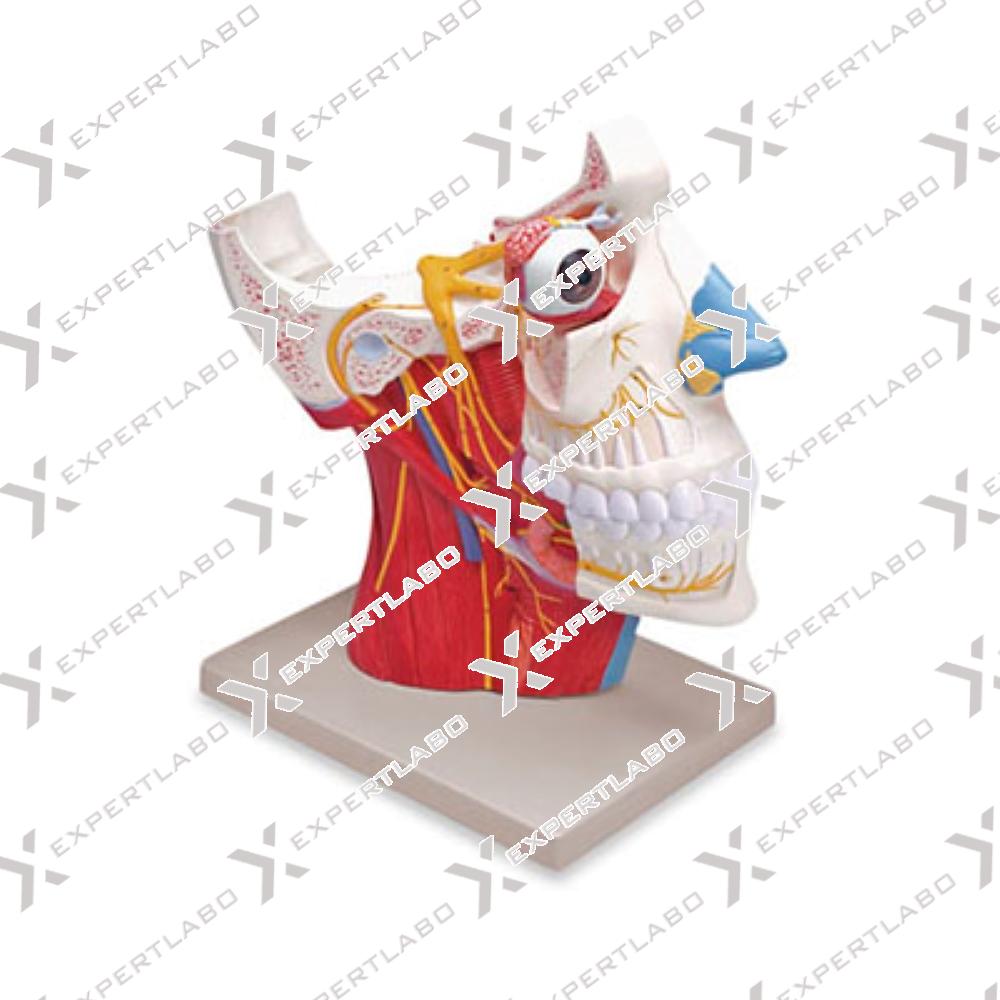
2.5X life size. Dissection of human half head shows the 12 cranial nerves with collateral branches and autonomic nerves. Median section reveals the upper respiratory tract and pharynx. The eyeball is removable for close examination. Mounted ...
View details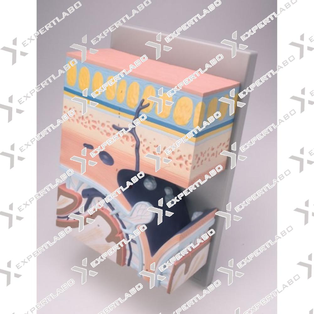
Enlarged several times. This model shows the stratification of the human cranium, revealing details of internal structures from the skin to the cerebral white matter. Mounted on board.
View details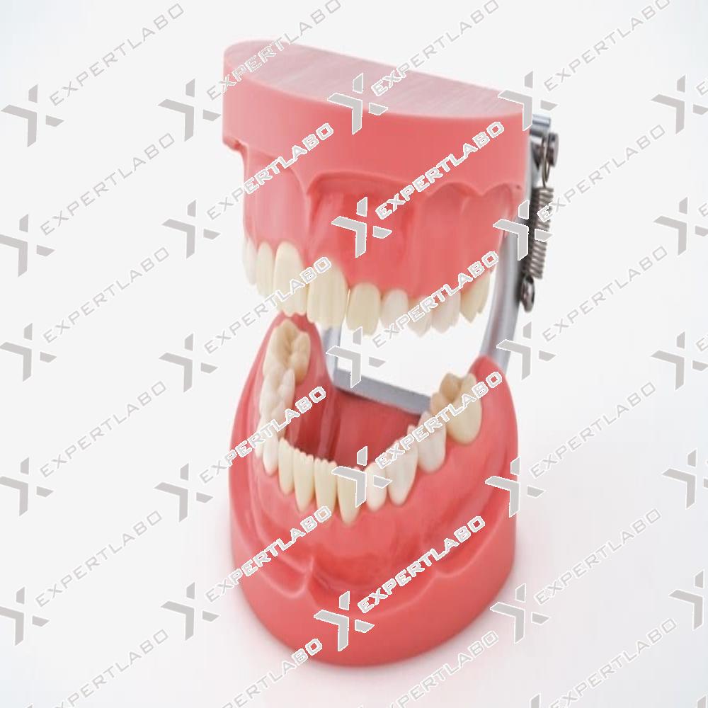
This 10-piece model is a useful tool to study the morphology of primary tooth types (incisors, canines and molars). Enlarged to10X life-size with mounted on board.
View details
A life size model representing the upper and lower jaw of an child, complete with deciduous and permanent teeth. The mandible is articulated to show natural range of movement when talking, breathing etc. Detailed underlying ...
View details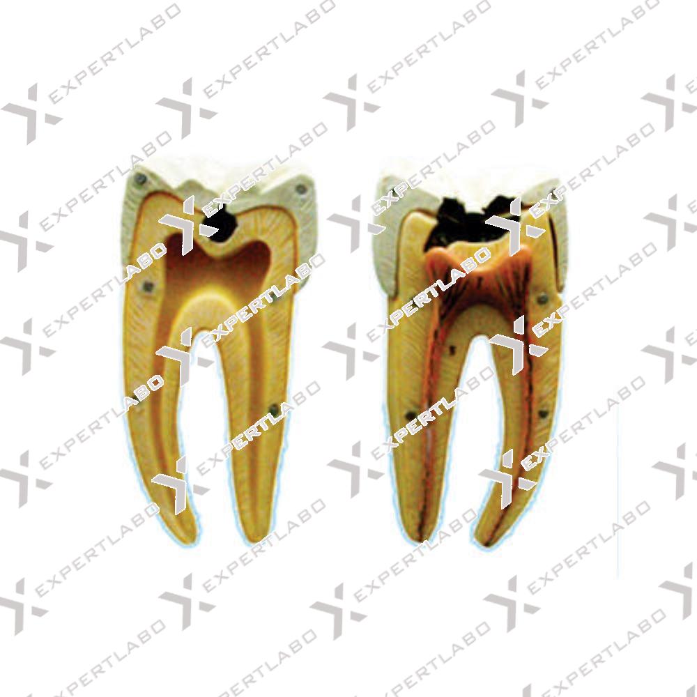
This model, composed of 7 parts, is 4X life size. It consists of 3 teeth, seated in their sockets, that shows the progressive deterioration of tooth structure by dental caries. The first tooth shows the caries in ...
View details
This life size model shows the upper and lower dentition of an adult human. Available aid for teaching proper oral hygiene procedures.
View details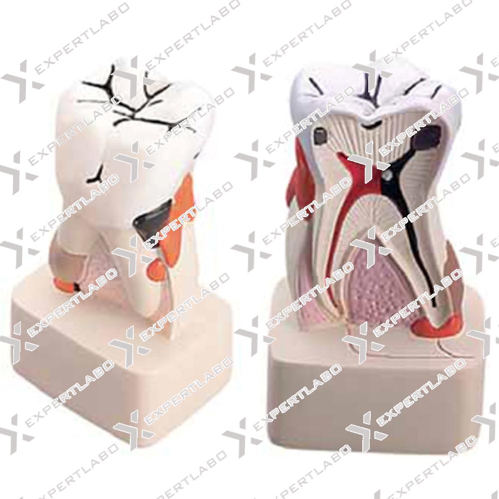
This 4X life size model shows the process of dental caries in its progressive stages. The tissues forming the tooth (enamel, dentine and pulp) are shown, along with the way in which they are affected ...
View details/1.jpg)
This model, enlarged 300X life size, shows the structure of a dicot wooden stem. Longitudinally and cross sectioned, it shows significant elements of the cortex and central vascular cylinder. All different structures are displayed in ...
View details/1.jpg)
This model, enlarged 300X life size, illustrates a dicot vascular structure. Longitudinally and cross sectioned, it shows clearly significant elements of the tegumental and cortical zone. The central vascular cylinder, including phloem and xylem, is ...
View details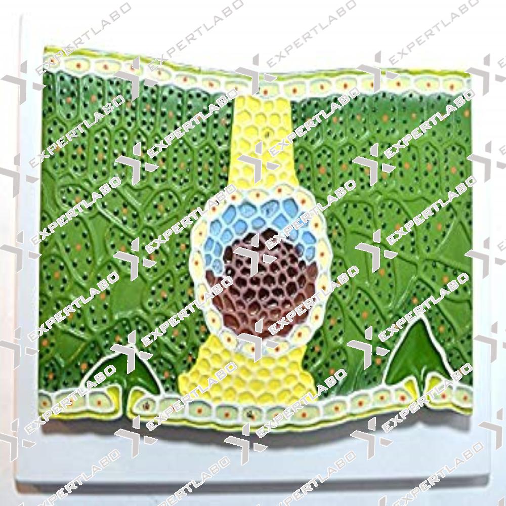
The structure of a dicot leaf is well represented in this 600X life-size model; the cross and the longitudinal section describes all the main structures and the different cellular types through the upper page, mesophyll ...
View details/1.jpg)
This 3-part model, enlarged 400X life size, shows the structure of a dicot root in a cross and longitudinal section. All significant structures are well represented in great detail. The model is divided in 3 segments ...
View details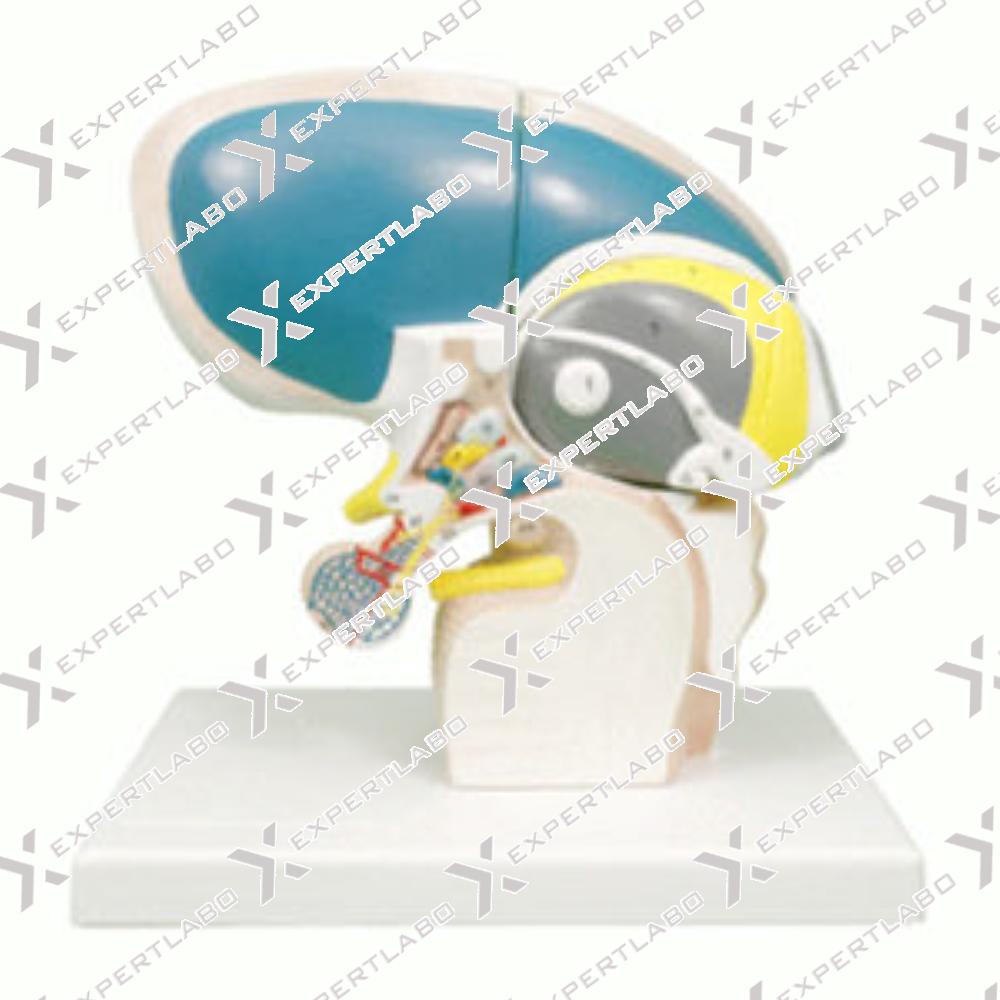
This 4-part model, 5X life-size, shows the human diencephalon: all the main parts of thalamus, epithalamus, metathalamus and hypothalamus are represented with great accuracy. The hypothalamic nuclei are shown in different colours; the thalamus is ...
View details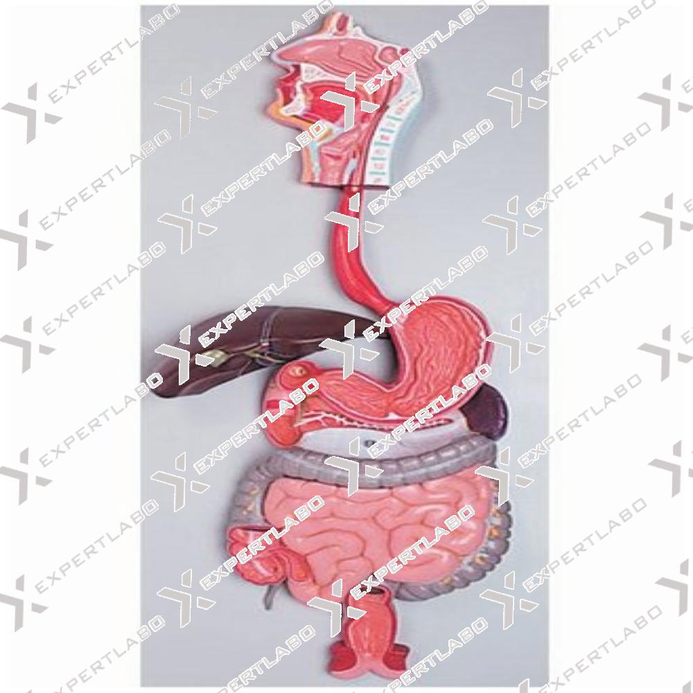
The life-size digestive system model demonstrates the entire digestive system in graphic relief. Nose, mouth cavity and pharynx, esophagus, Gl tract, liver with gall bladder, pancreas and spleen. The duodenum, caecum and rectum of the ...
View details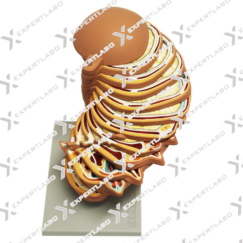
This life-size head model is sliced horizontally into 12 pieces, giving an idea of how computer tomography and magnetic resonance work. Each slice can be rotated and removed for closer examination of its anatomical detail, which ...
View details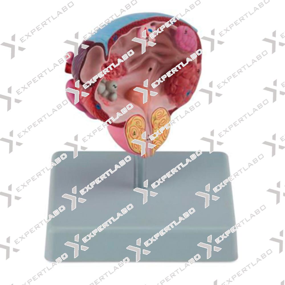
This life-size model, sectioned along the frontal plane, shows, in the appropriate location, five different pathologies of the male urinary bladder and prostate: Bladder stones, Cystitis, Diverticulum, Benign prostatic, Hypertrophy (BPH), Bladder tumour at three ...
View details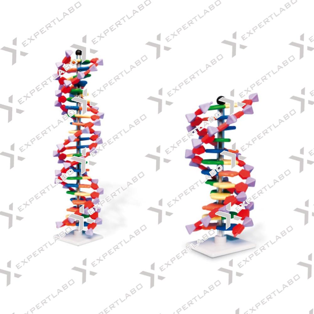
This self assembly kit contains all the blocks needed (stand included) to build a 12 base pairs right handed double helix DNA (B-DNA). Parts are colour coded and have characteristic shapes, with deoxyribose being pentagonal, phosphates ...
View details/1.jpg)
This life size model is an accurate representation of the pancreas, spleen and duodenum. The pancreas is open to show the entire pancreatic duct. The duodenum is partially dissected to expose its internal structure. Mounted ...
View details/1.jpg)
This model shows the effects of diabetes on eye, heart, kidney and foot The organs can be rotated for a detailed examination. Mounted on base with stand.
View details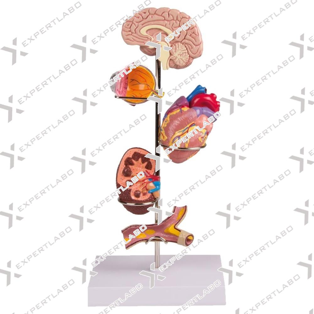
This model shows the effects of hypertension on brain, eye, heart, kidney and artery. The organs can be rotated for a detailed examination. Mounted on base with stand.
View details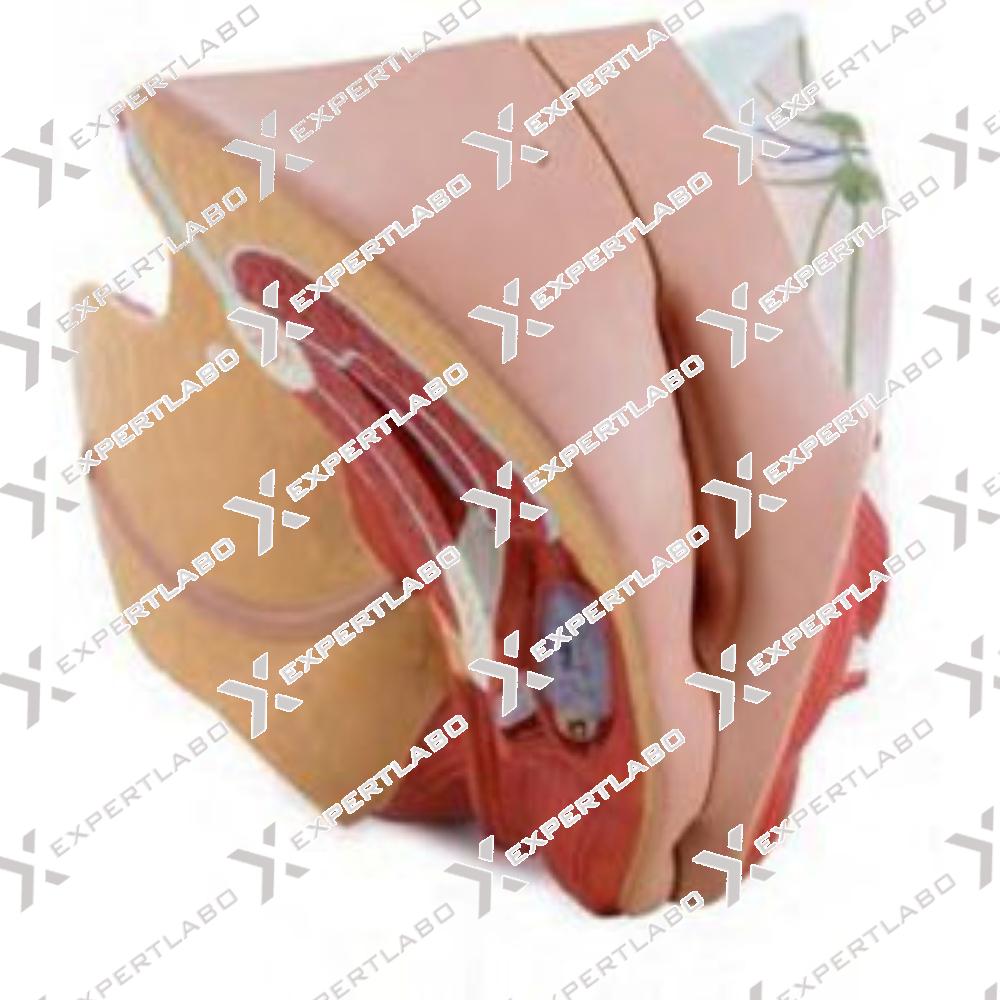
This life size model, composed of 4 parts, is a detailed representation of the female reproductive system as viewed through a median sagittal dissection. Removable parts include a 2-part uterus and 2 halves of the female reproductive ...
View details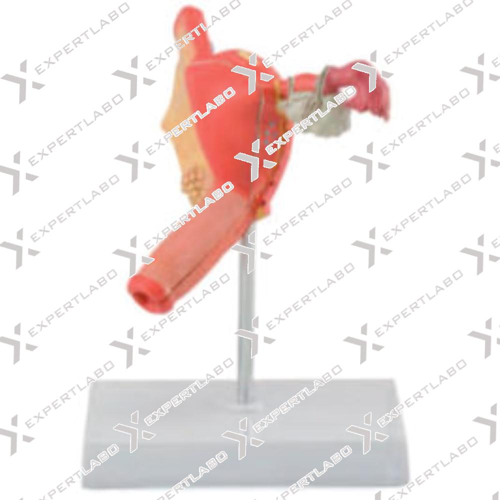
This life size model, sectioned along the front plane and divided into 2 pieces, shows the most important pathologies of the female reproductive system. Four types of fibroid tumors (intramural, subserous, submucous and broad ligament myoma) ...
View details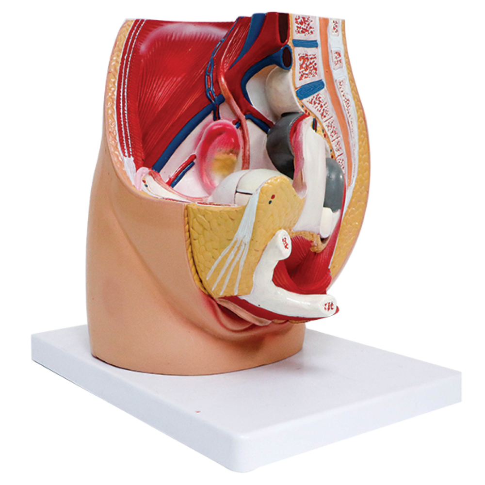
This single-piece life-size model is a very useful tool to study the female pelvic anatomy and to understand the relationship between pelvic bones, muscles and female genital organs. It shows a complete skeleton of a ...
View details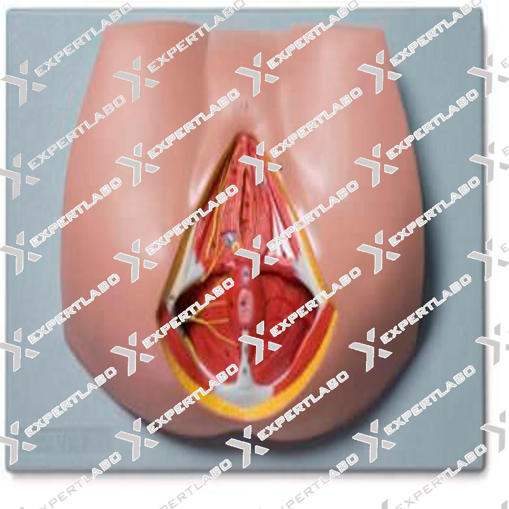
This life size model shows the female perineal area, including the anus and the external genitalia. The pelvic diaphragm, urogenital perineum with opening of the vagina and the anal perineum are well represented. Distribution of ...
View details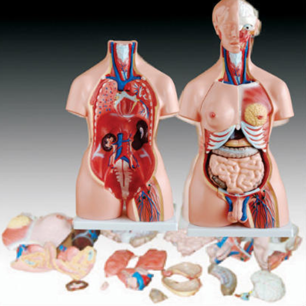
This female torso features 15 parts, including torso, head (2 parts), brain, lung (2 parts), heart (2 parts), stomach, liver, kidney, pancreas and spleen, intestine, female genitalia (2 parts). Made of fiber plastic. Mounted on plastic base.
View details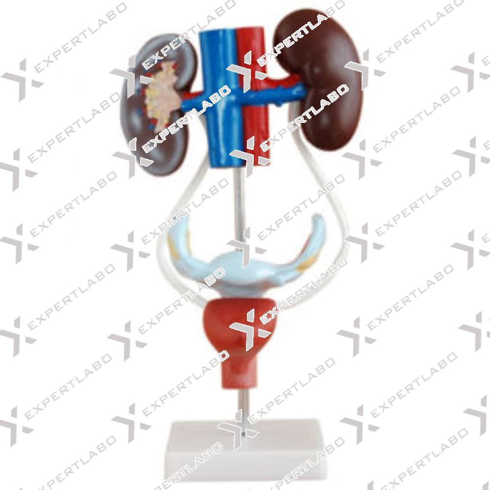
This model shows kidney, ureters, urinary bladder, uterus, accessories of uterus, vagina, ovary membrane, ligaments, uterus ligament and its artery etc. Made of PVC plastic with Mounted on stand. Refraction media: showing the lens and ...
View details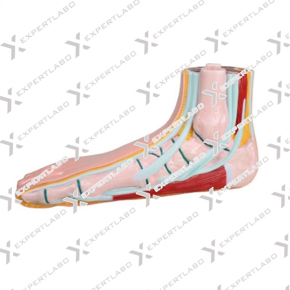
This life size model of a right human foot is medially opened and shows bones, joints, ligaments, fatty tissue and muscles. The start of lower leg is also represented as cross-section and shows tibia and ...
View details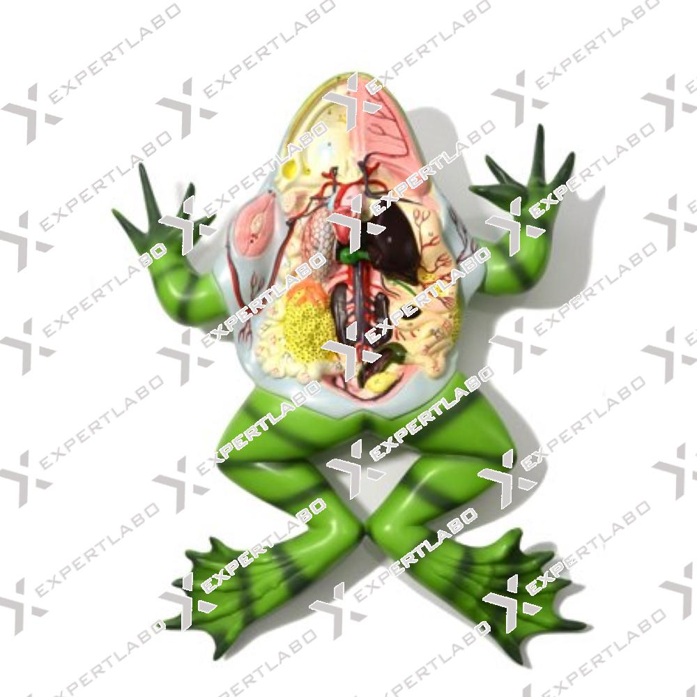
This 3X life size model provides a highly accurate illustration of the morphology of a frog. The ventral part is open for a closer study of the inner anatomy. Mounted on base with stand.
View details/1.jpg)
This 350X life size model shows in great detail the section of a pinus silvestris 3-year old stem. It describes all basic structures of the gymnosperm stem, including epidermis and vascular cylinder with mounted on ...
View details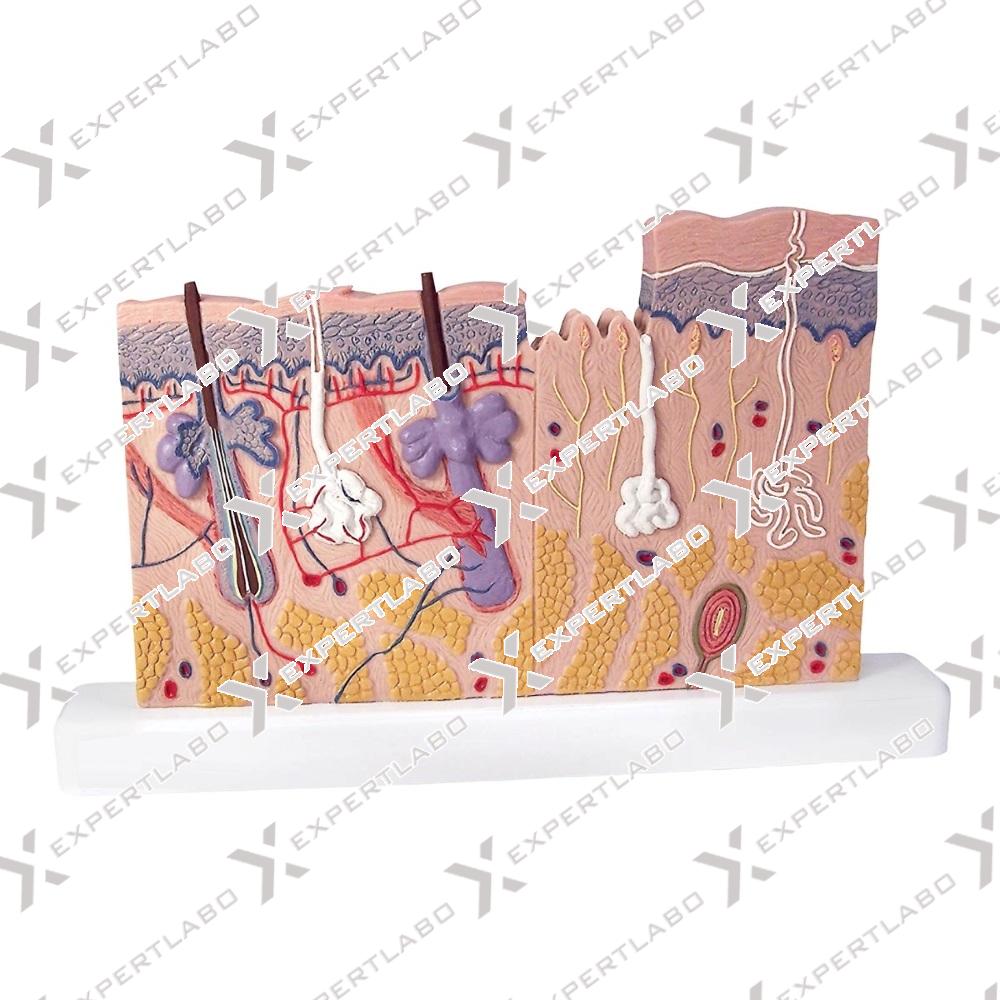
This 70X relief model compares a section of hairy skin to one without hair. All structures, including sensitive corpuscles, hair follicles, sebaceous and sweat glands, are shown in detail with Mounted on board.
View detailsCopyright © 2019. all rights reserved. Website designed by AV Web Solution
Expert Labo Has Regular Exports To The Following Countries: Afghanistan, Albania, Algeria, Andorra, Angola, Antigua And Barbuda, Argentina, Armenia, Australia, Austria, Azerbaijan, Bahamas, Bahrain, Bangladesh, Barbados, Belarus, Belgium, Belize, Benin, Bhutan, Bolivia, Bosnia And Herzegovina, Botswana, Brazil, Brunei, Bulgaria, Burkina Faso, Burma/ Myanmar, Burundi, Cambodia, Cameroon, Canada, Cape Verde, Central African Republic, Chad, Chile, Colombia, Comoros, Congo, Congo, Costa Rica, Cote D'Ivoire/Ivory Coast, Croatia, Cuba, Cyprus, Czech Republic, Denmark, Djibouti, Dominica, Dominican Republic, East Timor, Ecuador, Egypt, El Salvador, Equatorial Guinea, Eritrea, Estonia, Ethiopia (Addis Ababa), Fiji, Finland, France, Gabon, Gambia, Georgia, Germany, Ghana, Greece, Grenada, Guatemala, Guinea, Guinea-Bissau, Guyana, Haiti, Honduras, Hungary, Iceland, Indonesia, Iran, Iraq, Ireland, Israel, Italy, Jamaica, Japan, Jordan, Kazakstan, Kenya (Nairobi), Kiribati, Korea, North, Korea, South, Kuwait, Kyrgyzstan, Laos, Latvia, Lebanon, Lesotho, Liberia, Liechtenstein, Lithuania, Luxembourg, Macedonia, Madagascar, Malawi (Lilongwe), Malaysia (Kuala Lumpur), Maldives, Mali, Malta, Marshall Islands, Mauritania, Mauritius, Mexico, Micronesia, Moldova, Monaco, Mongolia, Montenegro, Morocco, Mozambique, Namibia, Nauru, Nepal, Netherlands, New Zealand, Nicaragua, Niger, Nigeria (Abuja), Norway, Oman, Palau, Panama, Papua New Guinea, Paraguay, Peru, Philippines (Manila), Poland, Portugal, Qatar, Romania, Russia, Rwanda (Kigali), Saint Kitts And Nevis, Saint Lucia, Saint Vincent And The Grenadines, Samoa, San Marino, Sao Tome And Principe, Saudi Arabia, Senegal, Serbia, Seychelles, Sierra Leone, Singapore, Slovakia, Slovenia, Solomon Islands, Somalia, South Africa, South Sudan, Spain, Sri Lanka, Sudan, Suriname, Swaziland, Sweden, Switzerland, Syria, Tajikistan, Tanzania, Thailand, Togo, Tonga, Trinidad And Tobago, Tunisia, Turkey, Turkmenistan, Tuvalu, Uganda (Kampala), Ukraine, United Arab Emirates (Dubai), United Kingdom (London), United States, Uruguay, Uzbekistan, Vanuatu, Venezuela, Vietnam, Yemen , Zambia (Lusaka), Zimbabwe